Department of Biomedical Engineering, Vanderbilt University, Nashville, Tennessee, USA.
Magn Reson Med. 2012 Dec;68(6):1774-84. doi: 10.1002/mrm.24186. Epub 2012 Jan 31.
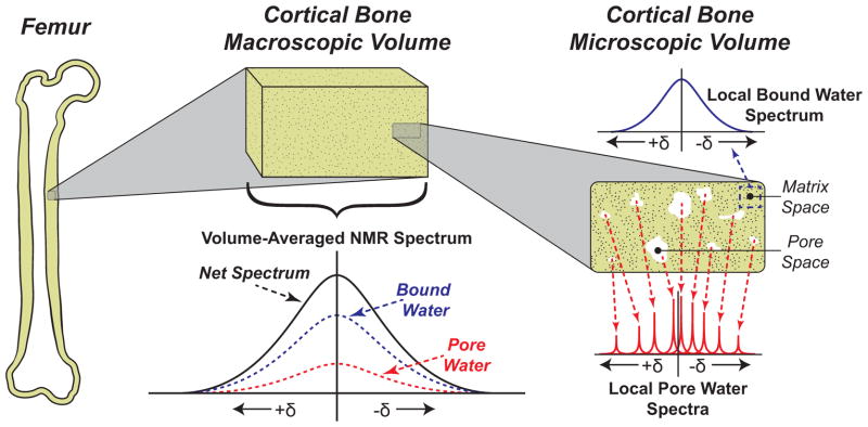
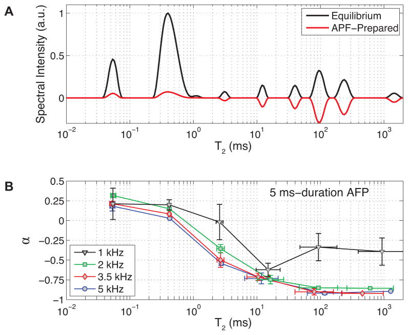
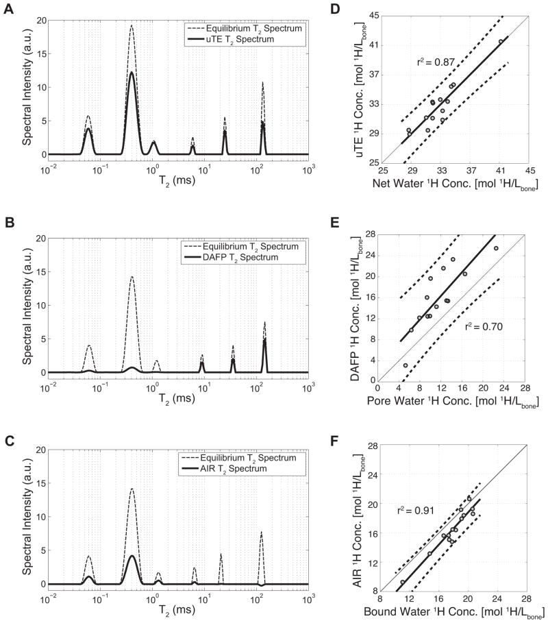
Advances in modern magnetic resonance imaging (MRI) pulse sequences have enabled clinically practical cortical bone imaging. Human cortical bone is known to contain a distribution of T(1) and T(2) components attributed to bound and pore water, although clinical imaging approaches have yet to discriminate bound from pore water based on their relaxation properties. Herein, two clinically compatible MRI strategies are proposed for selectively imaging either bound or pore water by utilizing differences in their T(1)s and T(2)s. The strategies are validated in a population of ex vivo human cortical bones, and estimates obtained for bound and pore water are compared to bone mechanical properties. Results show that the two MRI strategies provide good estimates of bound and pore water that correlate to bone mechanical properties. As such, the strategies for bound and pore water discrimination shown herein should provide diagnostically useful tools for assessing bone fracture risk, once applied to clinical MRI.
现代磁共振成像(MRI)脉冲序列的进步使得临床实用的皮质骨成像成为可能。已知人类皮质骨含有 T(1)和 T(2)分量的分布,这些分量归因于结合水和孔内水,尽管临床成像方法尚未根据其弛豫特性区分结合水和孔内水。本文提出了两种临床兼容的 MRI 策略,通过利用 T(1)和 T(2)的差异,选择性地对结合水或孔内水成像。这些策略在一组离体人皮质骨中进行了验证,并将获得的结合水和孔内水的估计值与骨力学性能进行了比较。结果表明,两种 MRI 策略都能很好地估计结合水和孔内水,且与骨力学性能相关。因此,一旦应用于临床 MRI,本文所示的用于区分结合水和孔内水的策略应该为评估骨折风险提供有用的诊断工具。