Universitätsklinik für Neurochirurgie, Medizinische Universität Graz, Austria.
Acta Neurochir (Wien). 2012 Apr;154(4):585-8; discussion 588. doi: 10.1007/s00701-012-1290-8.
Frameless stereotactic biopsies are replacing frame-based stereotaxy as a diagnostic approach to brain lesions. In order to avoid a sampling bias or negative histology, multiple specimens are often taken. This in turn increases the risk of hemorrhagic complications.
We present the use of 5-aminolevulinic acid (5-ALA)-induced protoporphyrin IX fluorescence in frameless stereotaxy to improve the procedure duration and yield, and thereby reduce the risk of complications.
Patients with suspected high-grade brain tumors are given 5-ALA 4 h prior to stereotactic biopsy. The biopsy needle is guided to the target using frameless stereotaxy based either on preoperative images or combined with intraoperative MRI sequences. The specimen is illuminated with blue light to look for fluorescence. In case of a positive fluorescence within the tissue sample, no frozen sections are obtained, and no further specimens are taken.
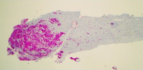
The samples of 13 patients revealed a positive fluorescence and were histologically confirmed as malignant or high-grade brain neoplasms. four cases were fluorescence-negative, requiring frozen section confirmation and/or multiple samples. In theses cases histology was either nonspecific gliotic changes or low-grade tumors. There were no complications related to the additional use of 5-ALA.
5-ALA fluorescence in stereotactic biopsies can increase the safety and accuracy of these procedures by reducing sampling errors and eliminating the need for multiple samples and/or frozen section verification, creating a more accurate, faster and safer procedure for cases of suspected malignant or high-grade brain tumors situated in deep or eloquent areas.
无框架立体定向活检正在取代框架立体定向术,成为脑病变的诊断方法。为了避免取样偏差或组织学阴性,通常会采集多个标本。这反过来又增加了出血并发症的风险。
我们介绍了在无框架立体定向术中使用 5-氨基酮戊酸(5-ALA)诱导的原卟啉 IX 荧光来改善手术过程的持续时间和产量,从而降低并发症的风险。
怀疑患有高级别脑肿瘤的患者在立体定向活检前 4 小时给予 5-ALA。活检针通过无框架立体定向术引导至目标,基于术前图像或结合术中 MRI 序列。用蓝光照射标本以寻找荧光。如果组织样本中有阳性荧光,则不获取冷冻切片,也不采集更多标本。
13 名患者的样本显示阳性荧光,组织学证实为恶性或高级别脑肿瘤。4 例为荧光阴性,需要冷冻切片确认和/或多个样本。这些病例的组织学表现为非特异性胶质改变或低级别肿瘤。没有与额外使用 5-ALA 相关的并发症。
5-ALA 荧光在立体定向活检中可以通过减少取样误差和消除对多个样本和/或冷冻切片验证的需求,提高这些手术的安全性和准确性,为位于深部或重要区域的疑似恶性或高级别脑肿瘤病例提供更准确、更快和更安全的手术。