Department of Respiratory Medicine, The First Affiliated Hospital of Xian Jiaotong University, School of Medicine, Xian, China.
Ann Thorac Med. 2012 Jan;7(1):21-5. doi: 10.4103/1817-1737.91559.
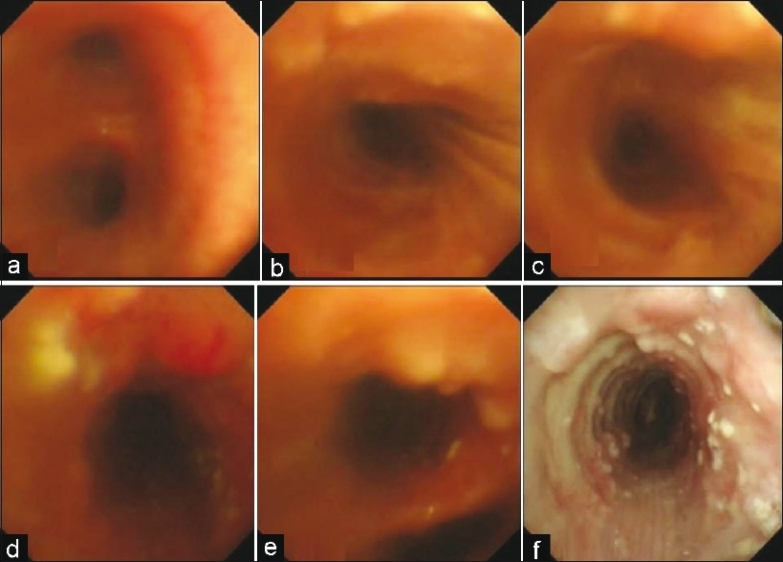
Auto-fluorescence bronchoscopy (AFB) has been used for the identification and localization of intra-epithelial pre-neoplastic and neoplastic lesions within the bronchus.
To determine the applicability of AFB for the detection and localization of precancerous and cancerous lesions, in addition to analyzing the morphologic presentation, their association to histological type and the variation between genders.
A five-year study involving 4983 patients, who underwent routine bronchoscopy [B] examination in a local tertiary teaching hospital, was done. The B examination was performed under intratracheal lidocaine, and samples were obtained using suitable approach. One thousand four hundred and eighty-five pathologically confirmed lung cancer patients were included in the study. The following parameters were studied: Morphological presentation, biopsy sites, histology. Differences between the groups were analyzed using Chi square test.
One thousand four hundred and eighty-five patients who had hyperplasia or neoplastic lesions were further confirmed as lung cancer pathologically. Lung cancer was more commonly found in the right lung (51.58% vs. 42.82%). The lesion occurred more frequently in the upper lobe than the lower lobe (44.17% vs. 22.42%). Male patients with squamous cell carcinoma showed upper lobe involvement more commonly, while the left main bronchus was more commonly involved in female patients. Adenocarcinoma mostly involved lesion of the upper lobe. Squamous cell carcinoma and small cell carcinoma were the major proliferative types (80.15% and 76.16% respectively).
AFB is efficient in the detection of pre-invasive and invasive lung lesions. The morphological presentation is associated to the histological type. There is variation in the presentation and histology of cancerous lung lesions between genders.
自动荧光支气管镜(AFB)已被用于识别和定位支气管内上皮前瘤变和肿瘤性病变。
确定 AFB 对癌前和癌性病变的检测和定位的适用性,同时分析形态表现、与组织学类型的关系以及性别之间的差异。
对一家地方三级教学医院进行常规支气管镜[B]检查的 4983 例患者进行了为期五年的研究。B 检查在气管内利多卡因下进行,通过合适的方法获取样本。1485 例经病理证实的肺癌患者纳入研究。研究的参数包括:形态表现、活检部位、组织学。使用卡方检验分析组间差异。
1485 例有增生或肿瘤性病变的患者进一步被病理证实为肺癌。右肺(51.58%比 42.82%)更常见肺癌。上叶比下叶更常发生病变(44.17%比 22.42%)。男性鳞状细胞癌患者更常出现上叶受累,而女性患者更常累及左主支气管。腺癌多累及上叶病变。鳞状细胞癌和小细胞癌是主要的增殖类型(分别为 80.15%和 76.16%)。
AFB 可有效检测肺前侵润和侵润性病变。形态表现与组织学类型有关。性别之间肺癌病变的表现和组织学存在差异。