• 文献检索
  • 文档翻译
  • 深度研究
  • 学术资讯
  • Suppr Zotero 插件Zotero 插件
  • 邀请有礼
  • 套餐&价格
  • 历史记录
应用&插件
Suppr Zotero 插件Zotero 插件浏览器插件Mac 客户端Windows 客户端微信小程序
定价
高级版会员购买积分包购买API积分包
服务
文献检索文档翻译深度研究API 文档MCP 服务
关于我们
关于 Suppr公司介绍联系我们用户协议隐私条款
关注我们

Suppr 超能文献

核心技术专利:CN118964589B侵权必究
粤ICP备2023148730 号-1Suppr @ 2026

文献检索

告别复杂PubMed语法,用中文像聊天一样搜索,搜遍4000万医学文献。AI智能推荐,让科研检索更轻松。

立即免费搜索

文件翻译

保留排版,准确专业,支持PDF/Word/PPT等文件格式,支持 12+语言互译。

免费翻译文档

深度研究

AI帮你快速写综述,25分钟生成高质量综述,智能提取关键信息,辅助科研写作。

立即免费体验

Flying through congested airspaces: imaging of chronic rhinosinusitis.

作者信息

Farina Davide, Ravanelli Marco, Borghesi Andrea, Maroldi Roberto

机构信息

Department of Radiology, University of Brescia, Piazza Spedali Civili 1, 25123 Brescia, Italy.

出版信息

Insights Imaging. 2010 Jul;1(3):155-166. doi: 10.1007/s13244-010-0030-y. Epub 2010 Jul 27.

DOI:10.1007/s13244-010-0030-y
PMID:22347913
原文链接:https://pmc.ncbi.nlm.nih.gov/articles/PMC3259331/
Abstract

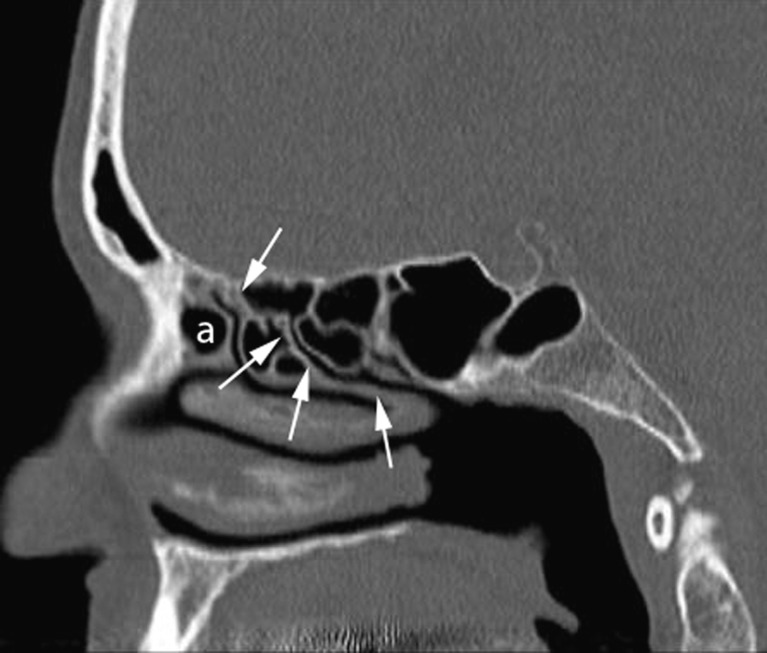
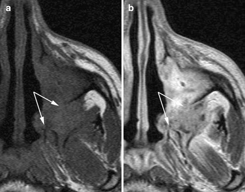
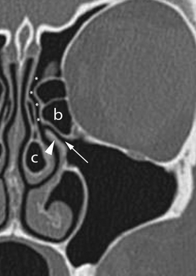
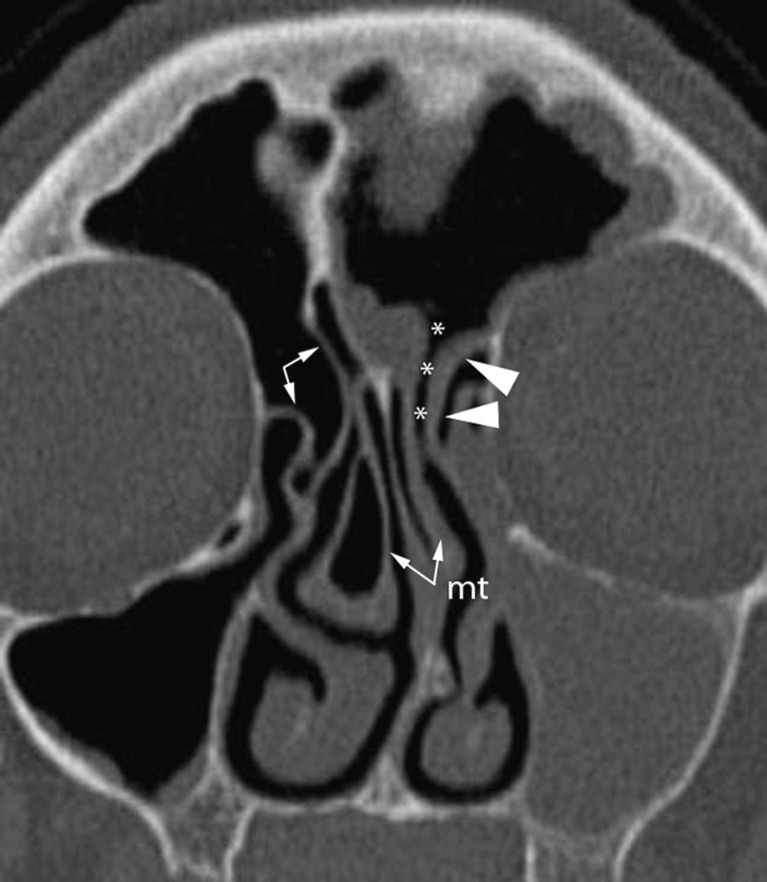
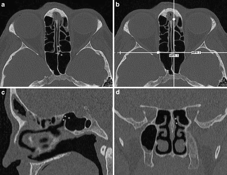
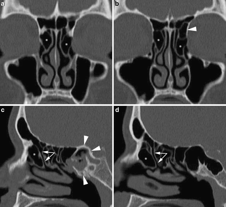
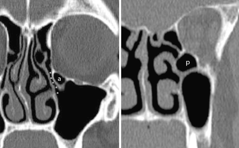
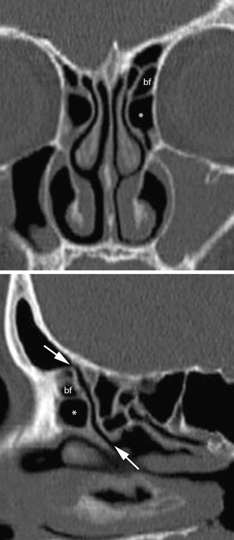
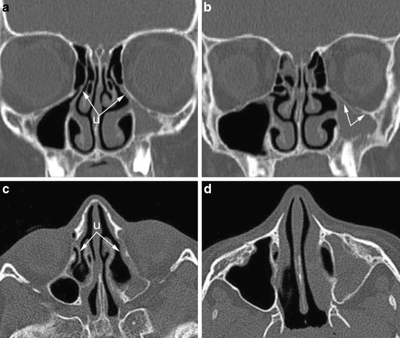
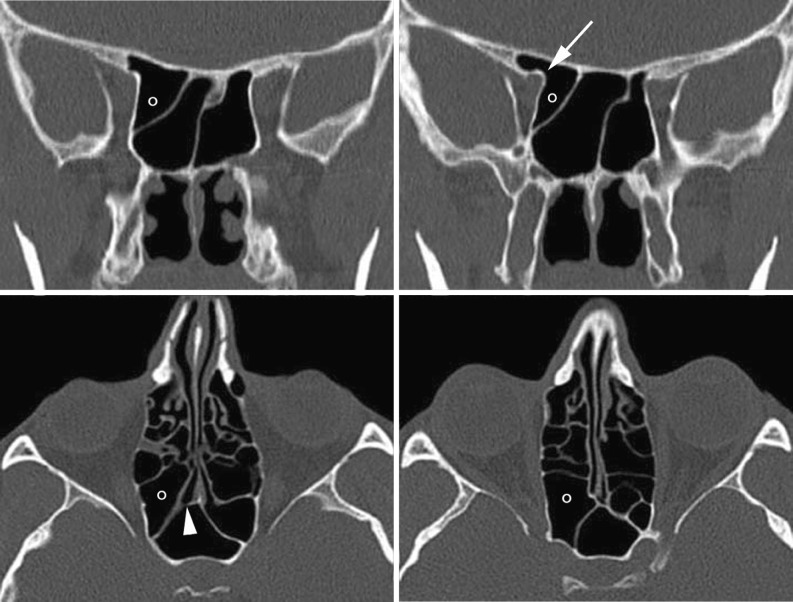
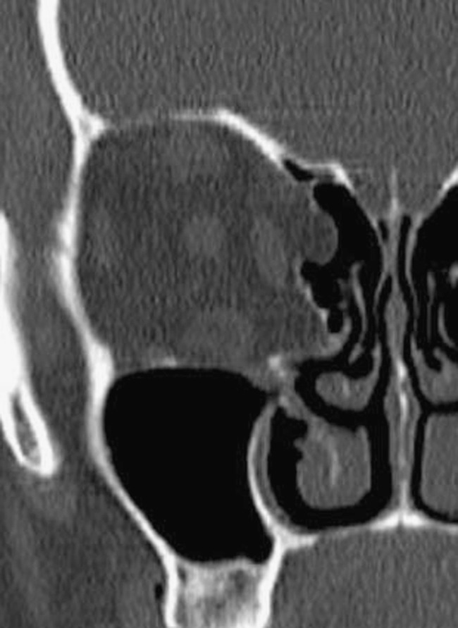
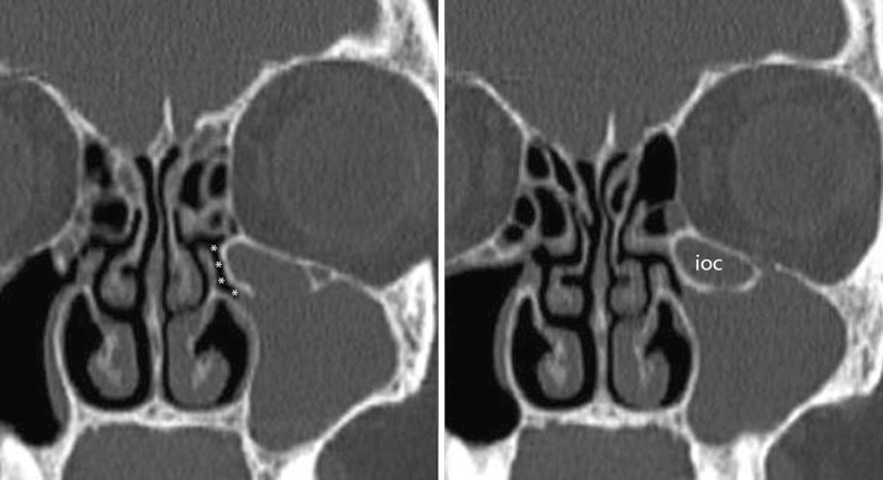
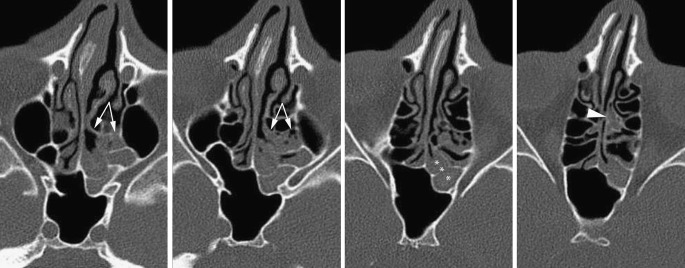
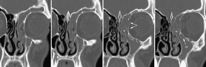
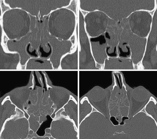
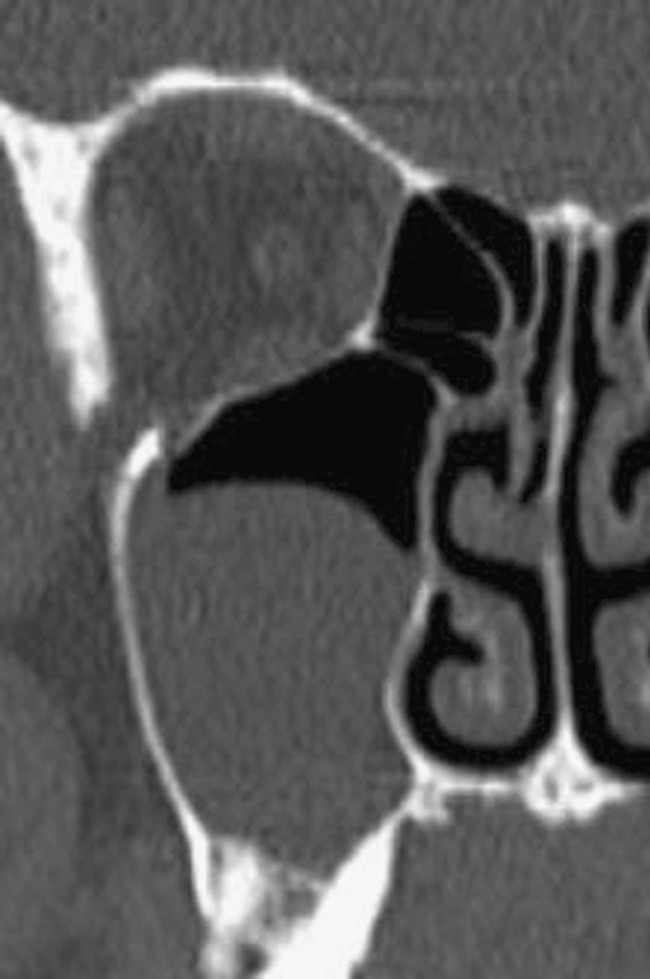
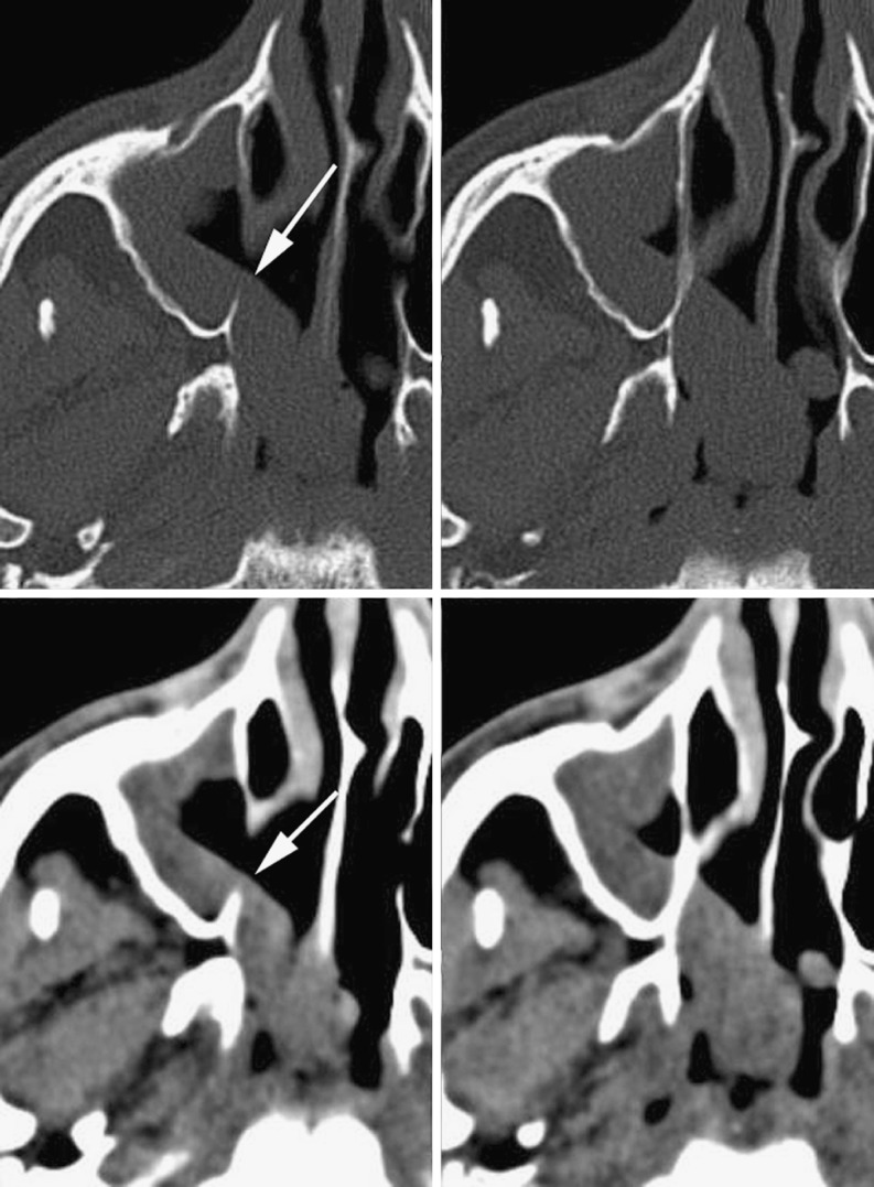
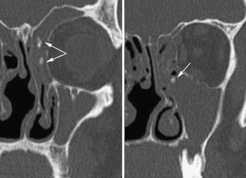
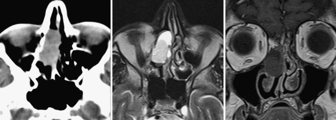
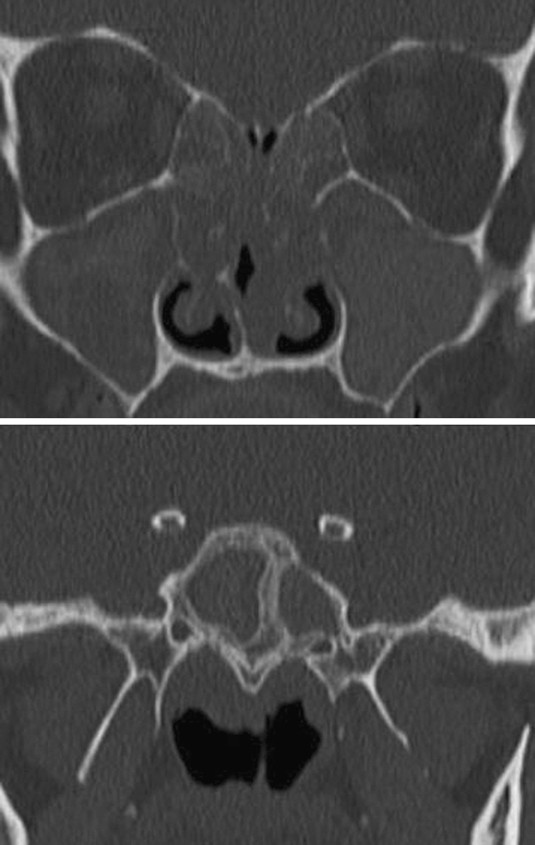
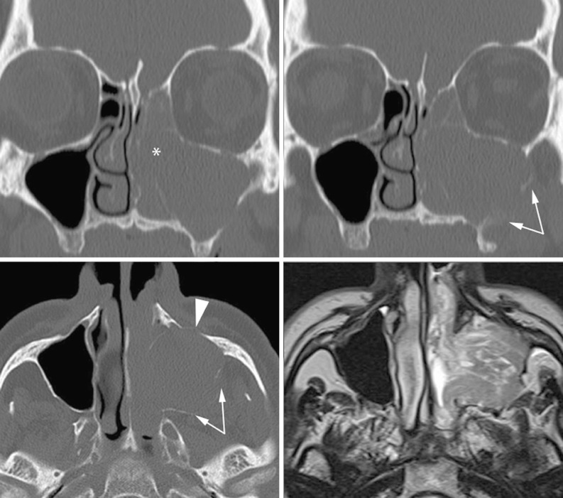
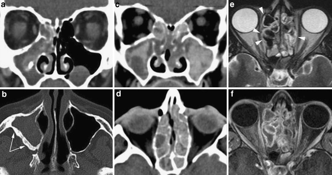
The complex regional anatomy of the nose and paranasal sinuses makes the interpretation of imaging studies of these structures intimidating to many radiologists. This paper aims to provide a key to interpretation by presenting a simplified approach to the functional anatomy of the paranasal sinuses and their most common (and most relevant) variants. This knowledge is basic for the full understanding of chronic rhinosinusitis and its computed tomography (CT) patterns. As fungal infections may be observed in the setting of chronic rhinosinusitis, these are also discussed. Chronic sinus inflammation produces bone changes, clearly depicted on CT images. Finally, clues to suspecting neoplastic lesions underlying inflammatory sinus conditions are provided.

摘要
https://cdn.ncbi.nlm.nih.gov/pmc/blobs/e108/3288852/b39792c2b618/13244_2010_30_Fig22_HTML.jpg
https://cdn.ncbi.nlm.nih.gov/pmc/blobs/e108/3288852/1834a08d3be4/13244_2010_30_Fig1_HTML.jpg
https://cdn.ncbi.nlm.nih.gov/pmc/blobs/e108/3288852/4ffdb91e9828/13244_2010_30_Fig2_HTML.jpg
https://cdn.ncbi.nlm.nih.gov/pmc/blobs/e108/3288852/3041c5d49695/13244_2010_30_Fig3_HTML.jpg
https://cdn.ncbi.nlm.nih.gov/pmc/blobs/e108/3288852/aac3ebe55785/13244_2010_30_Fig4_HTML.jpg
https://cdn.ncbi.nlm.nih.gov/pmc/blobs/e108/3288852/4bf0c1bfc944/13244_2010_30_Fig5_HTML.jpg
https://cdn.ncbi.nlm.nih.gov/pmc/blobs/e108/3288852/4edc1a9526fd/13244_2010_30_Fig6_HTML.jpg
https://cdn.ncbi.nlm.nih.gov/pmc/blobs/e108/3288852/247f13157e8f/13244_2010_30_Fig7_HTML.jpg
https://cdn.ncbi.nlm.nih.gov/pmc/blobs/e108/3288852/9cf6a950b032/13244_2010_30_Fig8_HTML.jpg
https://cdn.ncbi.nlm.nih.gov/pmc/blobs/e108/3288852/6a87e2d2546a/13244_2010_30_Fig9_HTML.jpg
https://cdn.ncbi.nlm.nih.gov/pmc/blobs/e108/3288852/ce8a9d990ad4/13244_2010_30_Fig10_HTML.jpg
https://cdn.ncbi.nlm.nih.gov/pmc/blobs/e108/3288852/3540dfc3d016/13244_2010_30_Fig11_HTML.jpg
https://cdn.ncbi.nlm.nih.gov/pmc/blobs/e108/3288852/ece4adc297da/13244_2010_30_Fig12_HTML.jpg
https://cdn.ncbi.nlm.nih.gov/pmc/blobs/e108/3288852/803cd09b4946/13244_2010_30_Fig13_HTML.jpg
https://cdn.ncbi.nlm.nih.gov/pmc/blobs/e108/3288852/2b2b70323eec/13244_2010_30_Fig14_HTML.jpg
https://cdn.ncbi.nlm.nih.gov/pmc/blobs/e108/3288852/4b4ab8dd1611/13244_2010_30_Fig15_HTML.jpg
https://cdn.ncbi.nlm.nih.gov/pmc/blobs/e108/3288852/e43cd5621255/13244_2010_30_Fig16_HTML.jpg
https://cdn.ncbi.nlm.nih.gov/pmc/blobs/e108/3288852/7e619bebdfd7/13244_2010_30_Fig17_HTML.jpg
https://cdn.ncbi.nlm.nih.gov/pmc/blobs/e108/3288852/27109e2abc84/13244_2010_30_Fig18_HTML.jpg
https://cdn.ncbi.nlm.nih.gov/pmc/blobs/e108/3288852/52bc1a112df3/13244_2010_30_Fig19_HTML.jpg
https://cdn.ncbi.nlm.nih.gov/pmc/blobs/e108/3288852/04cef744531e/13244_2010_30_Fig20_HTML.jpg
https://cdn.ncbi.nlm.nih.gov/pmc/blobs/e108/3288852/31ddf1e64988/13244_2010_30_Fig21_HTML.jpg
https://cdn.ncbi.nlm.nih.gov/pmc/blobs/e108/3288852/b39792c2b618/13244_2010_30_Fig22_HTML.jpg

相似文献

1
Flying through congested airspaces: imaging of chronic rhinosinusitis.
Insights Imaging. 2010 Jul;1(3):155-166. doi: 10.1007/s13244-010-0030-y. Epub 2010 Jul 27.
2
[Diseases of the nose and paranasal sinuses in childhood].[儿童鼻及鼻窦疾病]
Laryngorhinootologie. 2014 Mar;93 Suppl 1:S24-48. doi: 10.1055/s-0033-1363211. Epub 2014 Apr 7.
3
Computed Tomography in the Evaluation of Pathological Lesions of Paranasal Sinuses.计算机断层扫描在评估鼻窦病理病变中的应用
J Nepal Health Res Counc. 2015 May-Aug;13(30):116-20.
4
Functional anatomy and computed tomography imaging of the paranasal sinuses.鼻窦的功能解剖与计算机断层扫描成像
Am J Med Sci. 1998 Jul;316(1):2-12. doi: 10.1097/00000441-199807000-00002.
5
Paranasal sinus development and implications for imaging.鼻窦发育及其影像学意义。
Pediatr Radiol. 2021 Jun;51(7):1134-1148. doi: 10.1007/s00247-020-04859-y. Epub 2021 May 12.
6
Cone beam computed tomography for the nasal cavity and paranasal sinuses.用于鼻腔和鼻窦的锥形束计算机断层扫描
Dent Clin North Am. 2014 Jul;58(3):627-51. doi: 10.1016/j.cden.2014.04.003.
7
Prevalence of Anatomical Variations of the Sinonasal Region and their Relationship with Chronic Rhinosinusitis.鼻窦区域解剖变异的患病率及其与慢性鼻窦炎的关系。
Kathmandu Univ Med J (KUMJ). 2016;14(56):342-346.
8
Fungal rhinosinusitis.真菌性鼻-鼻窦炎
Prilozi. 2012;33(1):187-97.
9
Comparative Evaluation of Chronic Rhinosinusitis Patients by Conventional Radiography, Computed Tomography and Diagnostic Nasal Endoscopy (DNE).通过传统放射学、计算机断层扫描和诊断性鼻内镜检查(DNE)对慢性鼻窦炎患者进行比较评估。
Indian J Otolaryngol Head Neck Surg. 2016 Jun;68(2):173-8. doi: 10.1007/s12070-015-0958-9. Epub 2015 Dec 18.
10
Radiological imaging of inflammatory lesions in the nasal cavity and paranasal sinuses.鼻腔和鼻窦炎性病变的放射影像学检查
Eur Radiol. 2006 Apr;16(4):872-88. doi: 10.1007/s00330-005-0068-2. Epub 2006 Jan 4.

引用本文的文献

1
The Role of the Gut and Airway Microbiota in Chronic Rhinosinusitis with Nasal Polyps: A Systematic Review.肠道和气道微生物群在慢性鼻息肉鼻窦炎中的作用:系统评价。
Int J Mol Sci. 2024 Jul 27;25(15):8223. doi: 10.3390/ijms25158223.
2
Effect of photobiomodulation therapy on headache, and fatigue in patients with chronic rhinosinusitis: a randomized controlled study.光生物调节疗法对慢性鼻-鼻窦炎患者头痛和疲劳的影响:一项随机对照研究。
Lasers Med Sci. 2024 Feb 15;39(1):62. doi: 10.1007/s10103-024-04011-4.
3
An additional challenge for head and neck radiologists: anatomic variants posing a surgical risk - a pictorial review.

本文引用的文献

1
Antrochoanal polyp: analysis of 200 cases.上颌窦后鼻孔息肉:200例分析
Acta Otorhinolaryngol Ital. 2009 Feb;29(1):21-6.
2
[Sinonasal cavities: CT imaging features of anatomical variants and surgical risk].[鼻窦:解剖变异的CT成像特征与手术风险]
J Radiol. 2009 Jan;90(1 Pt 1):21-9. doi: 10.1016/s0221-0363(09)70074-5.
3
The development and pathologic processes that influence maxillary sinus pneumatization.影响上颌窦气化的发育及病理过程。
头颈放射科医生面临的另一个挑战:构成手术风险的解剖变异——图文综述。
Insights Imaging. 2019 Dec 2;10(1):112. doi: 10.1186/s13244-019-0794-7.
Anat Rec (Hoboken). 2008 Nov;291(11):1554-63. doi: 10.1002/ar.20774.
4
[Unilateral benign choanal polyp: review of 51 patients].[单侧良性后鼻孔息肉:51例患者的回顾]
Acta Otorrinolaringol Esp. 2008 Feb;59(2):52-6.
5
Imaging of chronic and exotic sinonasal disease: review.慢性及罕见鼻窦疾病的影像学:综述
AJR Am J Roentgenol. 2007 Dec;189(6 Suppl):S35-45. doi: 10.2214/AJR.07.7031.
6
Imaging features of invasive and noninvasive fungal sinusitis: a review.侵袭性和非侵袭性真菌性鼻窦炎的影像学特征:综述
Radiographics. 2007 Sep-Oct;27(5):1283-96. doi: 10.1148/rg.275065189.
7
Sphenochoanal polyp: an endoscopic view.蝶筛窦息肉:内镜视野
Ear Nose Throat J. 2007 Jan;86(1):19-20.
8
Radiological imaging of inflammatory lesions in the nasal cavity and paranasal sinuses.鼻腔和鼻窦炎性病变的放射影像学检查
Eur Radiol. 2006 Apr;16(4):872-88. doi: 10.1007/s00330-005-0068-2. Epub 2006 Jan 4.
9
The incidence of concha bullosa and its relationship to nasal septal deviation and paranasal sinus disease.泡状鼻甲的发病率及其与鼻中隔偏曲和鼻窦疾病的关系。
AJNR Am J Neuroradiol. 2004 Oct;25(9):1613-8.
10
3D computed tomographic analysis of frontal recess anatomy in patients without frontal sinusitis.无额窦炎患者额隐窝解剖结构的三维计算机断层扫描分析
Otolaryngol Head Neck Surg. 2004 Sep;131(3):164-73. doi: 10.1016/j.otohns.2004.04.012.