• 文献检索
  • 文档翻译
  • 深度研究
  • 学术资讯
  • Suppr Zotero 插件Zotero 插件
  • 邀请有礼
  • 套餐&价格
  • 历史记录
应用&插件
Suppr Zotero 插件Zotero 插件浏览器插件Mac 客户端Windows 客户端微信小程序
定价
高级版会员购买积分包购买API积分包
服务
文献检索文档翻译深度研究API 文档MCP 服务
关于我们
关于 Suppr公司介绍联系我们用户协议隐私条款
关注我们

Suppr 超能文献

核心技术专利:CN118964589B侵权必究
粤ICP备2023148730 号-1Suppr @ 2026

文献检索

告别复杂PubMed语法,用中文像聊天一样搜索,搜遍4000万医学文献。AI智能推荐,让科研检索更轻松。

立即免费搜索

文件翻译

保留排版,准确专业,支持PDF/Word/PPT等文件格式,支持 12+语言互译。

免费翻译文档

深度研究

AI帮你快速写综述,25分钟生成高质量综述,智能提取关键信息,辅助科研写作。

立即免费体验

克罗恩病中的超声造影:技术、图像解读及临床应用

Contrast-enhanced ultrasound (CEUS) in Crohn's disease: technique, image interpretation and clinical applications.

作者信息

Ripollés Tomás, Martínez-Pérez María J, Blanc Esther, Delgado Fructuoso, Vizuete José, Paredes José M, Vilar José

出版信息

Insights Imaging. 2011 Dec;2(6):639-652. doi: 10.1007/s13244-011-0124-1. Epub 2011 Aug 10.

DOI:10.1007/s13244-011-0124-1
PMID:22347983
原文链接:https://pmc.ncbi.nlm.nih.gov/articles/PMC3259412/
Abstract

BACKGROUND

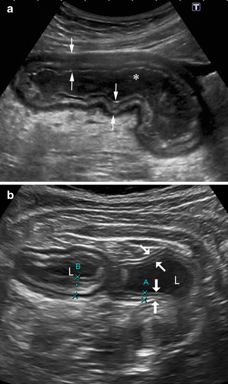
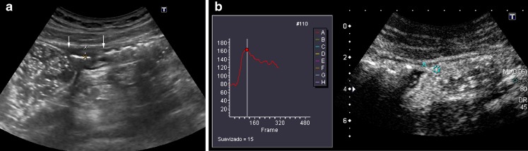
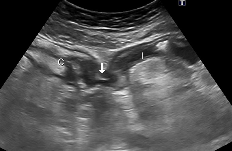
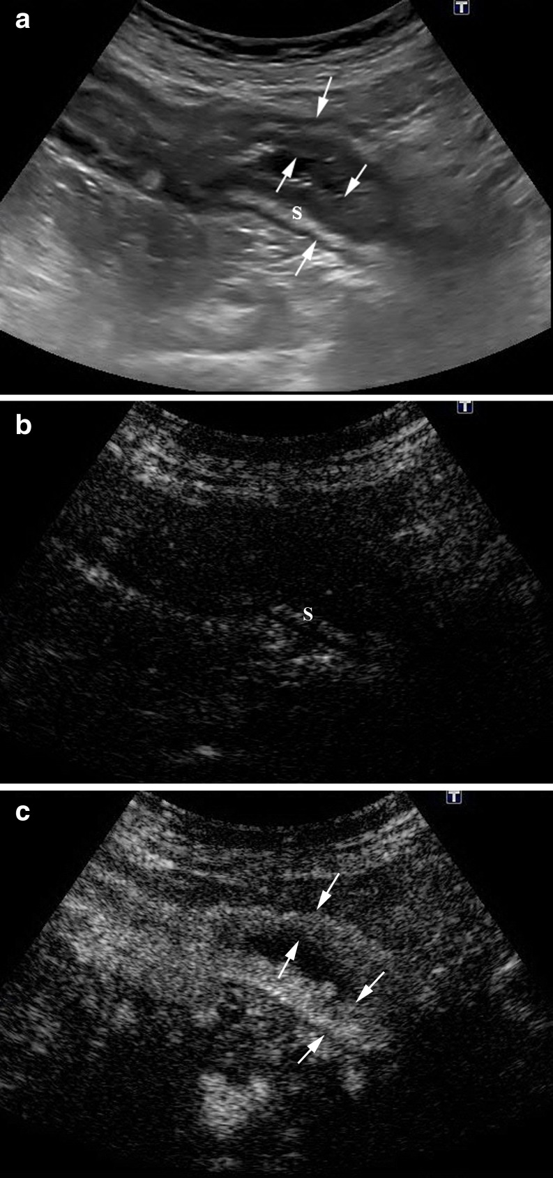
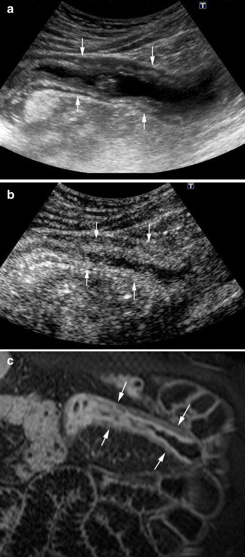
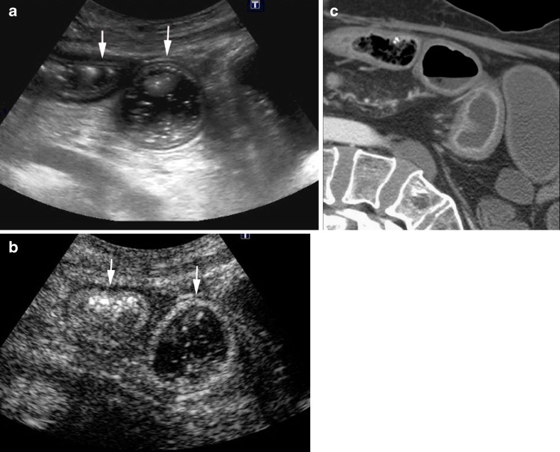
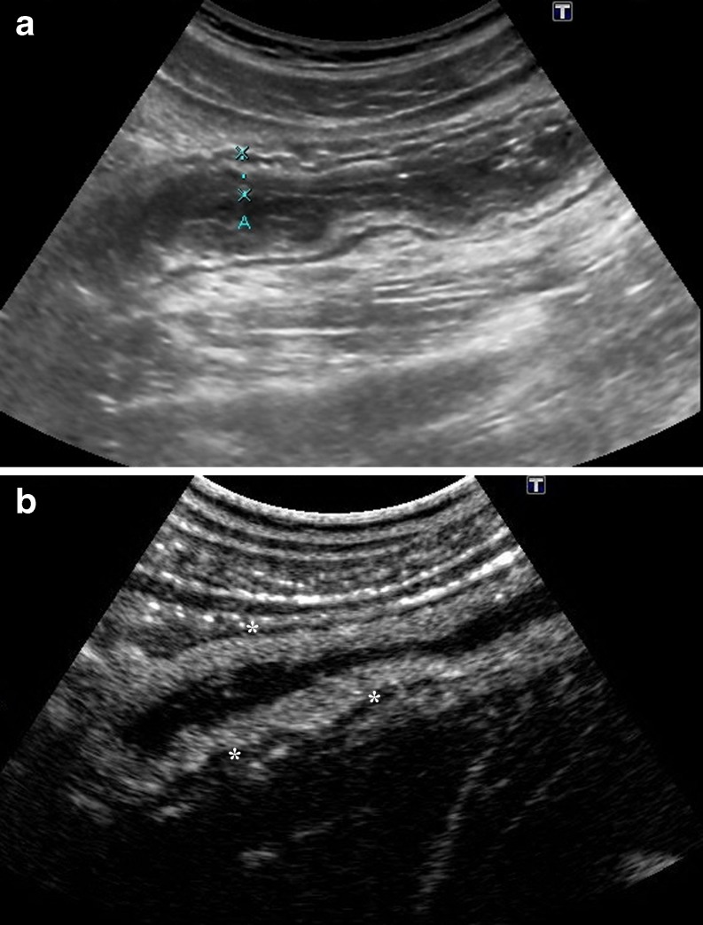
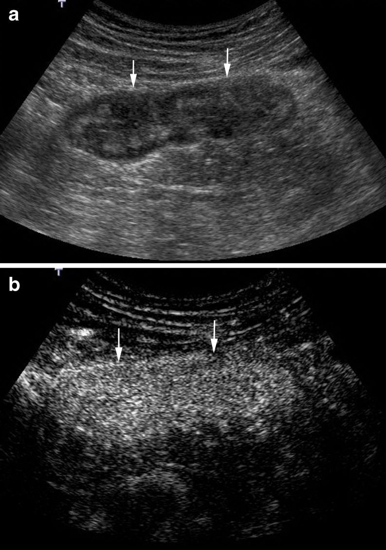
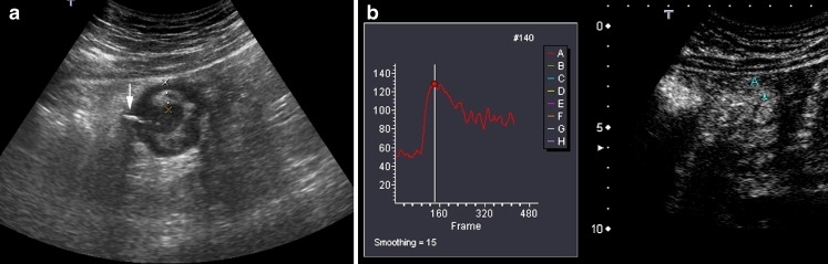
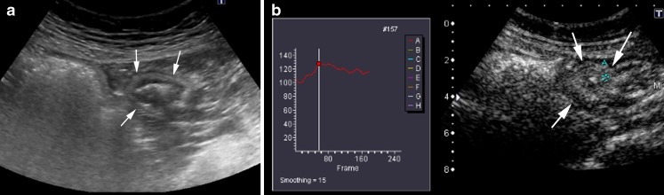
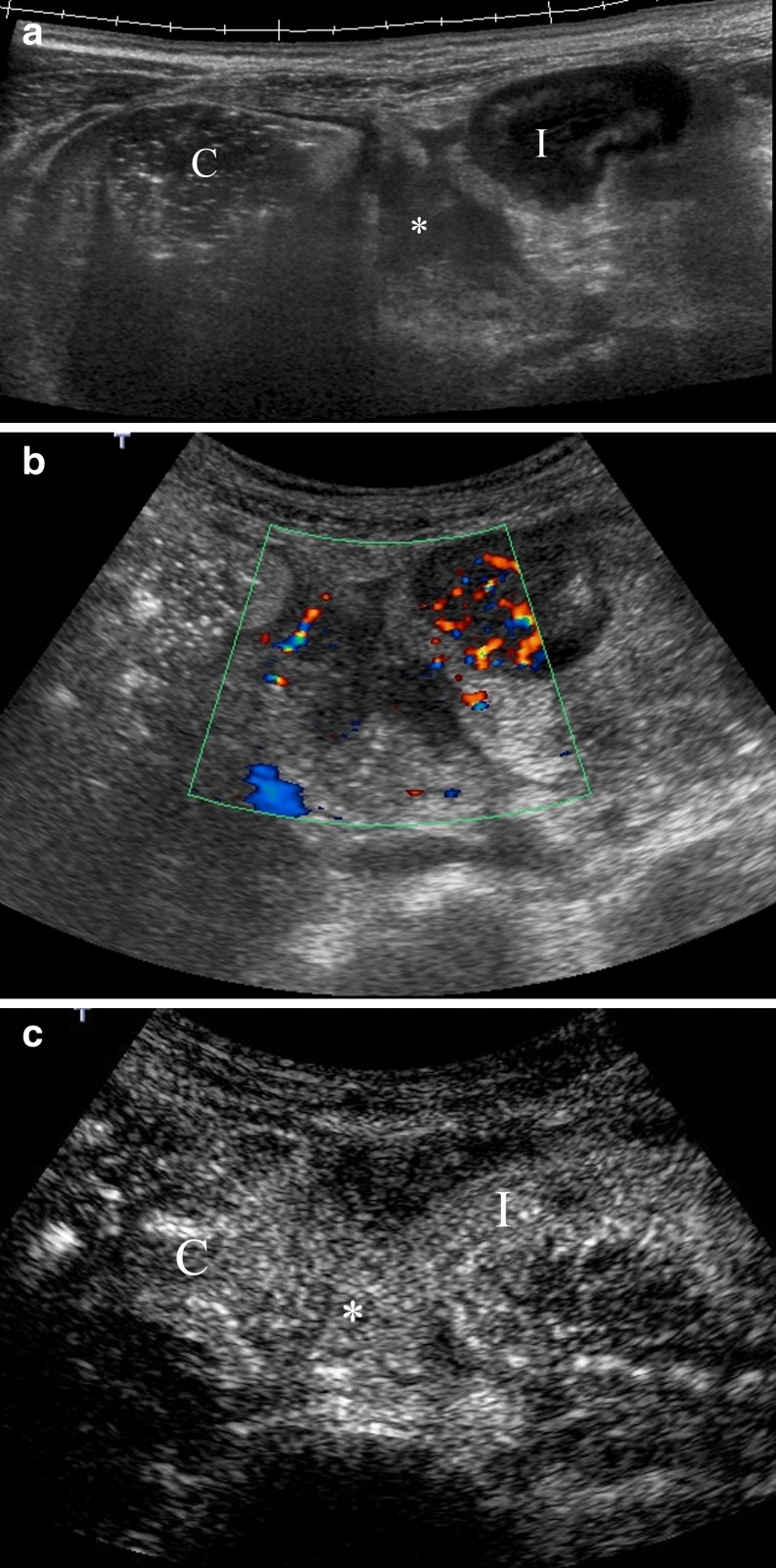
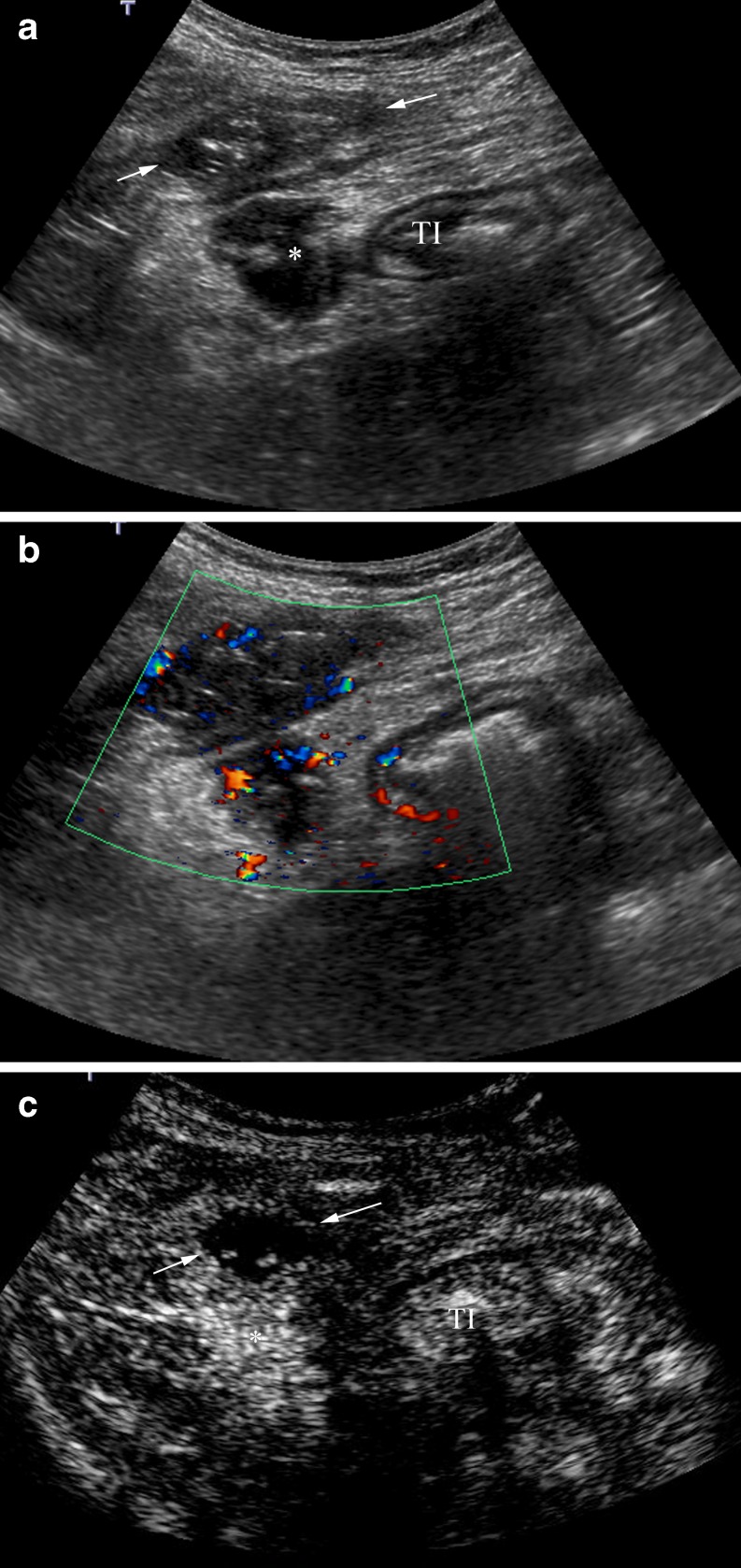
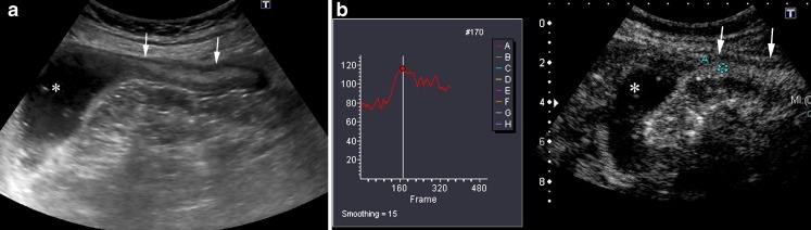
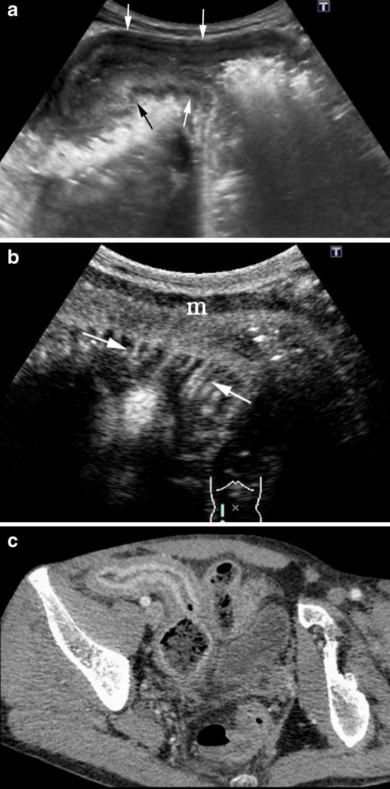
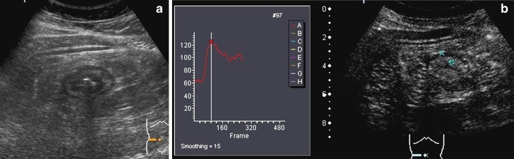
Recent meta-analysis has demonstrated no significant differences in diagnostic accuracy among different imaging techniques (US, MRI and CT) in the evaluation of Crohn's disease (CD). High-resolution bowel ultrasound has emerged as an alternative imaging technique for the diagnosis and follow-up of patients with CD, being as accurate as CT and MR for detecting intramural and extramural extension of the disease. B-Mode US can evaluate the localization and length of the affected intestinal segments and allow identification of transmural complications, stenosis and intestinal obstruction. Doppler techniques are tools that visualize and quantify bowel vascularization. Contrast-enhanced ultrasound (CEUS) is a new technique that involves IV administration of an ultrasound contrast agent with real-time examination, providing an accurate depiction of the bowel wall microvascularization and the perienteric tissues. The introduction of imaging quantification techniques enables an objective quantitative measurement of the enhancement. METHOD AND RESULTS: The article reviews the technique, sonographic findings, advantages and limitations, and clinical applications of contrast-enhanced US in the evaluation of Cohn's disease. Current CEUS applications in CD are: CD activity assessment, evaluation of inflammatory masses, distinguishing phlegmons from abscesses, characterization of stenosis by differentiating fibrosis from inflammation, monitoring the efficacy of drug treatments and improving the detection of disease recurrence. CONCLUSION: CEUS is an emerging technique that is part of the entire sonographic evaluation, with a role in the diagnosis and follow-up of CD, thus improving therapy planning and monitoring of the efficacy of treatment. ELECTRONIC SUPPLEMENTARY MATERIAL: The online version of this article (doi:10.1007/s13244-011-0124-1) contains supplementary material, which is available to authorized users.

摘要

背景

近期的荟萃分析表明,在克罗恩病(CD)评估中,不同成像技术(超声、磁共振成像和计算机断层扫描)的诊断准确性无显著差异。高分辨率肠道超声已成为CD患者诊断和随访的一种替代成像技术,在检测疾病的壁内和壁外扩展方面与CT和磁共振成像一样准确。B型超声可评估受累肠段的定位和长度,并有助于识别透壁并发症、狭窄和肠梗阻。多普勒技术是可视化和量化肠道血管化的工具。超声造影(CEUS)是一种新技术,涉及静脉注射超声造影剂并进行实时检查,可准确描绘肠壁微血管化和肠周组织。成像量化技术的引入能够对增强进行客观定量测量。

方法与结果

本文综述了超声造影在克罗恩病评估中的技术、超声表现、优缺点及临床应用。目前CEUS在CD中的应用包括:CD活动度评估、炎性肿块评估、鉴别蜂窝织炎与脓肿、通过区分纤维化与炎症来表征狭窄、监测药物治疗效果以及提高疾病复发的检测率。

结论

CEUS是一种新兴技术,是整个超声评估的一部分,在CD的诊断和随访中发挥作用,从而改善治疗方案规划和治疗效果监测。

电子补充材料

本文的在线版本(doi:https://doi.org/10.1007/s13244-011-0124-1)包含补充材料,授权用户可获取。 (注:原文中doi链接格式有误,补充材料获取部分根据正确格式添加了https:// ,实际翻译时可根据实际正确情况处理)

https://cdn.ncbi.nlm.nih.gov/pmc/blobs/4b86/3289034/b34999c80352/13244_2011_124_Fig18_HTML.jpg
https://cdn.ncbi.nlm.nih.gov/pmc/blobs/4b86/3289034/61c6df8ae970/13244_2011_124_Fig1_HTML.jpg
https://cdn.ncbi.nlm.nih.gov/pmc/blobs/4b86/3289034/901e3bc3c0b1/13244_2011_124_Fig2_HTML.jpg
https://cdn.ncbi.nlm.nih.gov/pmc/blobs/4b86/3289034/99fb1f554e05/13244_2011_124_Fig3_HTML.jpg
https://cdn.ncbi.nlm.nih.gov/pmc/blobs/4b86/3289034/4a7df068d5f6/13244_2011_124_Fig4_HTML.jpg
https://cdn.ncbi.nlm.nih.gov/pmc/blobs/4b86/3289034/f7c0cadbede0/13244_2011_124_Fig5_HTML.jpg
https://cdn.ncbi.nlm.nih.gov/pmc/blobs/4b86/3289034/45167ac22684/13244_2011_124_Fig6_HTML.jpg
https://cdn.ncbi.nlm.nih.gov/pmc/blobs/4b86/3289034/967ed43e8dd6/13244_2011_124_Fig7_HTML.jpg
https://cdn.ncbi.nlm.nih.gov/pmc/blobs/4b86/3289034/115e1c94ecc7/13244_2011_124_Fig8_HTML.jpg
https://cdn.ncbi.nlm.nih.gov/pmc/blobs/4b86/3289034/c4c879bd5e4c/13244_2011_124_Fig9_HTML.jpg
https://cdn.ncbi.nlm.nih.gov/pmc/blobs/4b86/3289034/7d88114b7b91/13244_2011_124_Fig10_HTML.jpg
https://cdn.ncbi.nlm.nih.gov/pmc/blobs/4b86/3289034/102b4ecf0861/13244_2011_124_Fig11_HTML.jpg
https://cdn.ncbi.nlm.nih.gov/pmc/blobs/4b86/3289034/adb19dfd9663/13244_2011_124_Fig12_HTML.jpg
https://cdn.ncbi.nlm.nih.gov/pmc/blobs/4b86/3289034/b809d76a006e/13244_2011_124_Fig13_HTML.jpg
https://cdn.ncbi.nlm.nih.gov/pmc/blobs/4b86/3289034/5d932fb0954e/13244_2011_124_Fig14_HTML.jpg
https://cdn.ncbi.nlm.nih.gov/pmc/blobs/4b86/3289034/017c128b0994/13244_2011_124_Fig15_HTML.jpg
https://cdn.ncbi.nlm.nih.gov/pmc/blobs/4b86/3289034/a0988b52cc9c/13244_2011_124_Fig16_HTML.jpg
https://cdn.ncbi.nlm.nih.gov/pmc/blobs/4b86/3289034/d4b80ae0b2c4/13244_2011_124_Fig17_HTML.jpg
https://cdn.ncbi.nlm.nih.gov/pmc/blobs/4b86/3289034/b34999c80352/13244_2011_124_Fig18_HTML.jpg

相似文献

1
Contrast-enhanced ultrasound (CEUS) in Crohn's disease: technique, image interpretation and clinical applications.克罗恩病中的超声造影:技术、图像解读及临床应用
Insights Imaging. 2011 Dec;2(6):639-652. doi: 10.1007/s13244-011-0124-1. Epub 2011 Aug 10.
2
Application of Dynamic Contrast-Enhanced Ultrasound in Evaluation the Activity of Crohn's Disease.动态对比增强超声在评估克罗恩病活动度中的应用
Diagnostics (Basel). 2024 Mar 22;14(7):672. doi: 10.3390/diagnostics14070672.
3
Contrast-enhanced ultrasonography (CEUS) vs. MRI of the small bowel in the evaluation of Crohn's disease activity.对比增强超声(CEUS)与 MRI 小肠成像在评估克罗恩病活动度中的比较。
Radiol Med. 2012 Mar;117(2):268-81. doi: 10.1007/s11547-011-0783-5. Epub 2012 Jan 21.
4
Assessment of the extension and the inflammatory activity in Crohn's disease: comparison of ultrasound and MRI.克罗恩病中病变范围及炎症活性的评估:超声与磁共振成像的比较
Abdom Imaging. 2009 Mar-Apr;34(2):141-8. doi: 10.1007/s00261-008-9365-y.
5
Effectiveness of contrast-enhanced ultrasound for characterisation of intestinal inflammation in Crohn's disease: a comparison with surgical histopathology analysis.对比增强超声在克罗恩病肠道炎症特征描述中的有效性:与手术组织病理学分析的比较。
J Crohns Colitis. 2013 Mar;7(2):120-8. doi: 10.1016/j.crohns.2012.03.002. Epub 2012 Apr 5.
6
The Role of Intravenous Contrast Agent in the Sonographic Assessment of Crohn's Disease Activity: Is Contrast Agent Injection Necessary?静脉造影剂在克罗恩病活动的超声评估中的作用:是否有必要注射造影剂?
J Crohns Colitis. 2019 Apr 26;13(5):585-592. doi: 10.1093/ecco-jcc/jjy204.
7
Dynamic contrast-enhanced ultrasound of the bowel wall with quantitative assessment of Crohn's disease activity in childhood.儿童克罗恩病活动度定量评估的肠壁动态对比增强超声检查
Radiol Oncol. 2016 Nov 9;50(4):347-354. doi: 10.1515/raon-2015-0042. eCollection 2016 Dec 1.
8
Contrast enhanced ultrasound: comparing a novel modality to MRI to assess for bowel disease in pediatric Crohn's patients.对比增强超声:将一种新型检查方法与磁共振成像对比,以评估儿童克罗恩病患者的肠道疾病
Transl Gastroenterol Hepatol. 2020 Apr 5;5:13. doi: 10.21037/tgh.2019.11.02. eCollection 2020.
9
Contrast-enhanced ultrasound of the small bowel in Crohn's disease.克罗恩病中小肠的对比增强超声检查
Abdom Imaging. 2013 Oct;38(5):1005-13. doi: 10.1007/s00261-013-0014-8.
10
Predictive value of time-intensity curves obtained with contrast-enhanced ultrasonography (CEUS) in the follow-up of 30 patients with Crohn's disease.对比增强超声(CEUS)获得的时间-强度曲线在30例克罗恩病患者随访中的预测价值
J Ultrasound. 2009 Dec;12(4):151-9. doi: 10.1016/j.jus.2009.10.001. Epub 2009 Nov 5.

引用本文的文献

1
Magnetic Resonance Enterography and Intestinal Ultrasound for the Assessment and Monitoring of Crohn's Disease.磁共振肠道成像和肠道超声用于评估和监测克罗恩病。
J Crohns Colitis. 2024 Sep 3;18(9):1450-1463. doi: 10.1093/ecco-jcc/jjae042.
2
Diagnostic Accuracy of Intestinal Ultrasound in the Detection of Intra-Abdominal Complications in Crohn's Disease: A Systematic Review and Meta-Analysis.肠超声诊断克罗恩病腹腔内并发症的准确性:系统评价和荟萃分析。
J Crohns Colitis. 2024 Jun 3;18(6):958-972. doi: 10.1093/ecco-jcc/jjad215.
3
Crohn's Disease: Basic Characteristics of the Disease, Diagnostic Methods, the Role of Biomarkers, and Analysis of Metalloproteinases: A Review.

本文引用的文献

1
Systematic review: the use of ultrasonography, computed tomography and magnetic resonance imaging for the diagnosis, assessment of activity and abdominal complications of Crohn's disease.系统评价:超声、计算机断层扫描和磁共振成像在克罗恩病的诊断、活动评估及腹部并发症中的应用。
Aliment Pharmacol Ther. 2011 Jul;34(2):125-45. doi: 10.1111/j.1365-2036.2011.04710.x. Epub 2011 May 25.
2
Non-invasive diagnosis and grading of postsurgical endoscopic recurrence in Crohn's disease: usefulness of abdominal ultrasonography and (99m)Tc-hexamethylpropylene amineoxime-labelled leucocyte scintigraphy.经内镜手术后克罗恩病复发的非侵入性诊断和分级:腹部超声和(99m)Tc-六甲基丙烯酰胺肟标记白细胞闪烁显像的作用。
J Crohns Colitis. 2010 Nov;4(5):537-45. doi: 10.1016/j.crohns.2010.03.002. Epub 2010 Apr 8.
3
克罗恩病:疾病的基本特征、诊断方法、生物标志物的作用以及金属蛋白酶分析:综述
Life (Basel). 2023 Oct 15;13(10):2062. doi: 10.3390/life13102062.
4
Integrating Intestinal Ultrasound into an Inflammatory Bowel Disease Practice: How to Get Started.将肠道超声纳入炎症性肠病诊疗实践:如何起步。
Crohns Colitis 360. 2023 Aug 7;5(3):otad043. doi: 10.1093/crocol/otad043. eCollection 2023 Jul.
5
Submucosal fat accumulation in Crohn's disease: evaluation with sonography.克罗恩病黏膜下脂肪堆积:超声评估
Intest Res. 2023 Jul;21(3):385-391. doi: 10.5217/ir.2022.00030. Epub 2023 Mar 17.
6
Aptamer-Functionalized Microbubbles Targeted to P-selectin for Ultrasound Molecular Imaging of Murine Bowel Inflammation.靶向P-选择素的适体功能化微泡用于小鼠肠道炎症的超声分子成像
Mol Imaging Biol. 2023 Apr;25(2):283-293. doi: 10.1007/s11307-022-01755-9. Epub 2022 Jul 18.
7
Ultrasonographic scores for ileal Crohn's disease assessment: Better, worse or the same as contrast-enhanced ultrasound?超声评分用于评估回肠克罗恩病:优于、劣于还是等同于对比增强超声?
BMC Gastroenterol. 2022 May 18;22(1):252. doi: 10.1186/s12876-022-02326-6.
8
Current and Emerging Approaches to the Diagnosis and Treatment of Crohn's Disease Strictures.克罗恩病狭窄诊断与治疗的当前及新出现的方法
Gastroenterol Hepatol (N Y). 2022 Apr;18(4):186-195.
9
Contrast-enhanced ultrasonography in dogs with inflammatory bowel disease.对比增强超声在犬炎症性肠病中的应用。
J Vet Intern Med. 2021 Sep;35(5):2167-2176. doi: 10.1111/jvim.16202. Epub 2021 Aug 25.
10
Contrast-enhanced ultrasound of the pediatric bowel.小儿肠的超声造影检查。
Pediatr Radiol. 2021 Nov;51(12):2214-2228. doi: 10.1007/s00247-020-04868-x. Epub 2021 May 12.
Which magnetic resonance imaging findings accurately evaluate inflammation in small bowel Crohn's disease? A retrospective comparison with surgical pathologic analysis.哪些磁共振成像发现能准确评估小肠克罗恩病的炎症?与手术病理分析的回顾性比较。
Inflamm Bowel Dis. 2011 Apr;17(4):984-93. doi: 10.1002/ibd.21414. Epub 2010 Aug 18.
4
Crohn disease: correlation of findings at contrast-enhanced US with severity at endoscopy.克罗恩病:对比增强超声检查结果与内镜检查严重程度的相关性
Radiology. 2009 Oct;253(1):241-8. doi: 10.1148/radiol.2531082269. Epub 2009 Jul 27.
5
Contrast-enhanced ultrasonographic evaluation of inflammatory activity in Crohn's disease.克罗恩病炎症活动的超声造影评估
Gastroenterology. 2009 Jul;137(1):43-52. doi: 10.1053/j.gastro.2009.03.062. Epub 2009 May 5.
6
Abdominal sonographic changes after antibody to tumor necrosis factor (anti-TNF) alpha therapy in Crohn's Disease.抗肿瘤坏死因子-α(anti-TNF-α)治疗克罗恩病后的腹部超声变化。
Dig Dis Sci. 2010 Feb;55(2):404-10. doi: 10.1007/s10620-009-0759-7. Epub 2009 Mar 7.
7
The diagnostic value of small bowel wall vascularity after sulfur hexafluoride-filled microbubble injection in patients with Crohn's disease. Correlation with the therapeutic effectiveness of specific anti-inflammatory treatment.六氟化硫填充微泡注射后小肠壁血管情况对克罗恩病患者的诊断价值。与特异性抗炎治疗疗效的相关性。
Eur J Radiol. 2009 Mar;69(3):438-44. doi: 10.1016/j.ejrad.2008.10.029. Epub 2008 Dec 12.
8
Inflammatory bowel disease diagnosed with US, MR, scintigraphy, and CT: meta-analysis of prospective studies.超声、磁共振成像、闪烁扫描法及计算机断层扫描诊断炎症性肠病:前瞻性研究的荟萃分析
Radiology. 2008 Apr;247(1):64-79. doi: 10.1148/radiol.2471070611.
9
Inflammatory activity in Crohn disease: ultrasound findings.克罗恩病的炎症活动:超声检查结果
Abdom Imaging. 2008 Sep-Oct;33(5):589-97. doi: 10.1007/s00261-007-9340-z.
10
Crohn's disease and color Doppler sonography: response to treatment and its relationship with long-term prognosis.克罗恩病与彩色多普勒超声检查:对治疗的反应及其与长期预后的关系。
J Clin Ultrasound. 2008 Jun;36(5):267-72. doi: 10.1002/jcu.20423.