Department of Morphology, Institute of Biological Sciences, Federal University of Minas Gerais, Belo Horizonte, Brazil.
J Gynecol Oncol. 2012 Jan;23(1):11-5. doi: 10.3802/jgo.2012.23.1.11. Epub 2012 Jan 9.
This study focused on comparing the expression levels of p16, Ki-67, and minichromosome maintenance 7 (MCM7) protein in normal and affected cervical epithelium to ascertain the biological significance of these markers in detecting progressive cervical disease.
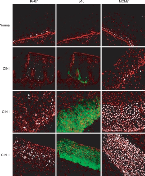
A quantitative and based on-scanning-microscopy analysis of the three markers expression was performed in normal and cervical intraepithelial neoplasia (CIN) I, II, and III tissues. p16 area as well as p16, Ki-67, and MCM7 positive cells or nuclei were evaluated according to their distribution and extent through the cervical epithelium.
A clear p16 over-expression was observed in all the dysplastic epithelium tissue samples. The quantitative analysis of p16 area as well as the number of p16 positive cells was able to better discriminate the CIN lesions grades than the usual semi-quantitative analysis. The average Ki-67 labeling indexes for the normal epithelium, CIN I, CIN II, and CIN III groups were 19.8%, 27.3%, 32.8%, and 37.1%, respectively, whereas the mean MCM7 labeling indexes for the correspondent grades were 27.0%, 30.4%, 50.5%, and 67.2%. The Ki-67 and MCM7 labeling indexes were closely correlated with the CIN histological grade, with higher labeling indexe values obtained from the more severe lesions (p<0.05), being the MCM7 labeling indexes the highest values in all the CIN categories (p<0.05).
We observed a good correlation among the p16, Ki-67, and MCM7 data. In addition, MCM7 demonstrated to be a more efficient and sensitive marker to assess disease progression in the uterine cervix.
本研究旨在比较正常和病变宫颈上皮中 p16、Ki-67 和微小染色体维持蛋白 7(MCM7)的表达水平,以确定这些标志物在检测宫颈疾病进展中的生物学意义。
采用定量和基于扫描显微镜的分析方法,检测正常和宫颈上皮内瘤变(CIN)I、II、III 组织中这三种标志物的表达。根据 p16、Ki-67 和 MCM7 阳性细胞或核在宫颈上皮中的分布和程度,评估 p16 面积以及 p16、Ki-67 和 MCM7 阳性细胞或核的数量。
所有发育不良的上皮组织样本中均观察到明显的 p16 过表达。p16 面积的定量分析以及 p16 阳性细胞的数量能够更好地区分 CIN 病变的分级,优于通常的半定量分析。正常上皮、CIN I、CIN II 和 CIN III 组的平均 Ki-67 标记指数分别为 19.8%、27.3%、32.8%和 37.1%,而相应分级的平均 MCM7 标记指数分别为 27.0%、30.4%、50.5%和 67.2%。Ki-67 和 MCM7 标记指数与 CIN 组织学分级密切相关,病变越严重,标记指数越高(p<0.05),其中 MCM7 标记指数在所有 CIN 分级中最高(p<0.05)。
我们观察到 p16、Ki-67 和 MCM7 数据之间存在良好的相关性。此外,MCM7 被证明是评估子宫颈疾病进展的更有效和敏感的标志物。