Department of Neuroradiology, Medical Faculty Mannheim, University of Heidelberg, Mannheim, Germany.
PLoS One. 2012;7(2):e31179. doi: 10.1371/journal.pone.0031179. Epub 2012 Feb 8.
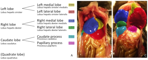
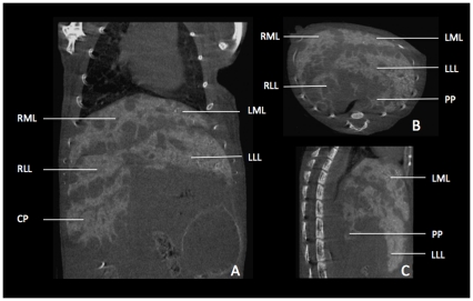
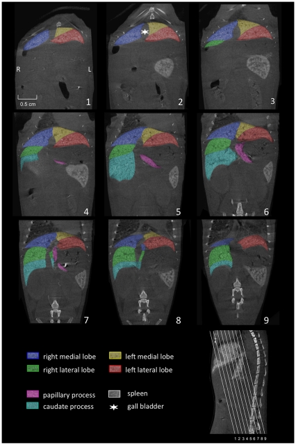
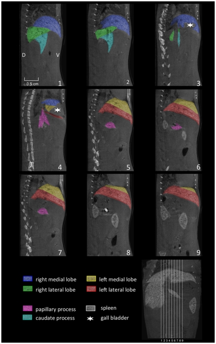
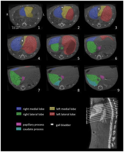
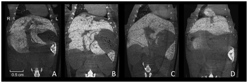
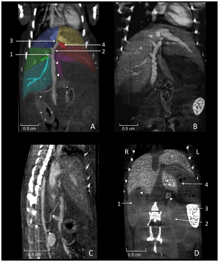
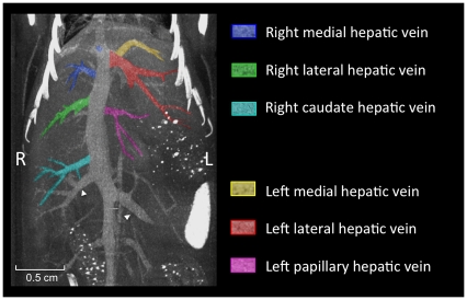
Various murine models are currently used to study acute and chronic pathological processes of the liver, and the efficacy of novel therapeutic regimens. The increasing availability of high-resolution small animal imaging modalities presents researchers with the opportunity to precisely identify and describe pathological processes of the liver. To meet the demands, the objective of this study was to provide a three-dimensional illustration of the macroscopic anatomical location of the murine liver lobes and hepatic vessels using small animal imaging modalities. We analysed micro-CT images of the murine liver by integrating additional information from the published literature to develop comprehensive illustrations of the macroscopic anatomical features of the murine liver and hepatic vasculature. As a result, we provide updated three-dimensional illustrations of the macroscopic anatomy of the murine liver and hepatic vessels using micro-CT. The information presented here provides researchers working in the field of experimental liver disease with a comprehensive, easily accessable overview of the macroscopic anatomy of the murine liver.
目前,多种鼠类模型被用于研究肝脏的急性和慢性病理过程,以及新型治疗方案的疗效。高分辨率小动物成像技术的日益普及为研究人员提供了精确识别和描述肝脏病理过程的机会。为了满足这一需求,本研究的目的是使用小动物成像技术对鼠类肝脏叶和肝血管的宏观解剖位置进行三维描绘。我们通过整合来自已发表文献的其他信息来分析鼠类肝脏的微计算机断层扫描图像,从而开发出鼠类肝脏和肝血管宏观解剖特征的综合示意图。结果,我们使用微计算机断层扫描提供了鼠类肝脏和肝血管宏观解剖的更新三维示意图。这里提供的信息为从事实验性肝病研究的研究人员提供了鼠类肝脏宏观解剖的全面、易于获取的概述。