• 文献检索
  • 文档翻译
  • 深度研究
  • 学术资讯
  • Suppr Zotero 插件Zotero 插件
  • 邀请有礼
  • 套餐&价格
  • 历史记录
应用&插件
Suppr Zotero 插件Zotero 插件浏览器插件Mac 客户端Windows 客户端微信小程序
定价
高级版会员购买积分包购买API积分包
服务
文献检索文档翻译深度研究API 文档MCP 服务
关于我们
关于 Suppr公司介绍联系我们用户协议隐私条款
关注我们

Suppr 超能文献

核心技术专利:CN118964589B侵权必究
粤ICP备2023148730 号-1Suppr @ 2026

文献检索

告别复杂PubMed语法,用中文像聊天一样搜索,搜遍4000万医学文献。AI智能推荐,让科研检索更轻松。

立即免费搜索

文件翻译

保留排版,准确专业,支持PDF/Word/PPT等文件格式,支持 12+语言互译。

免费翻译文档

深度研究

AI帮你快速写综述,25分钟生成高质量综述,智能提取关键信息,辅助科研写作。

立即免费体验

人脑头皮脑电图中传播性去极化的相关因素。

Correlates of spreading depolarization in human scalp electroencephalography.

机构信息

Centre for Stroke Research Berlin, Charité University Medicine Berlin, 10117 Berlin, Germany.

出版信息

Brain. 2012 Mar;135(Pt 3):853-68. doi: 10.1093/brain/aws010.

DOI:10.1093/brain/aws010
PMID:22366798
原文链接:https://pmc.ncbi.nlm.nih.gov/articles/PMC3286336/
Abstract

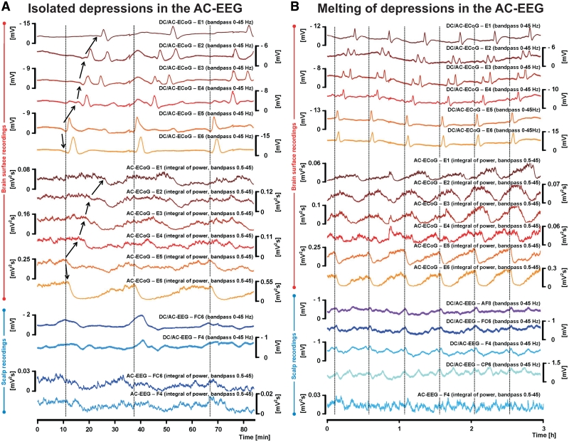
It has been known for decades that suppression of spontaneous scalp electroencephalographic activity occurs during ischaemia. Trend analysis for such suppression was found useful for intraoperative monitoring during carotid endarterectomy, or as a screening tool to detect delayed cerebral ischaemia after aneurismal subarachnoid haemorrhage. Nevertheless, pathogenesis of such suppression of activity has remained unclear. In five patients with aneurismal subarachnoid haemorrhage and four patients with decompressive hemicraniectomy after malignant hemispheric stroke due to middle cerebral artery occlusion, we here performed simultaneously full-band direct and alternating current electroencephalography at the scalp and direct and alternating current electrocorticography at the cortical surface. After subarachnoid haemorrhage, 275 slow potential changes, identifying spreading depolarizations, were recorded electrocorticographically over 694 h. Visual inspection of time-compressed scalp electroencephalography identified 193 (70.2%) slow potential changes [amplitude: -272 (-174, -375) µV (median quartiles), duration: 5.4 (4.0, 7.1) min, electrocorticography-electroencephalography delay: 1.8 (0.8, 3.5) min]. Intervals between successive spreading depolarizations were significantly shorter for depolarizations with electroencephalographically identified slow potential change [33.0 (27.0, 76.5) versus 53.0 (28.0, 130.5) min, P = 0.009]. Electroencephalography was thus more likely to display slow potential changes of clustered than isolated spreading depolarizations. In contrast to electrocorticography, no spread of electroencephalographic slow potential changes was seen, presumably due to superposition of volume-conducted electroencephalographic signals from widespread cortical generators. In two of five patients with subarachnoid haemorrhage, serial magnetic resonance imaging revealed large delayed infarcts at the recording site, while electrocorticography showed clusters of spreading depolarizations with persistent depression of spontaneous activity. Alternating current electroencephalography similarly displayed persistent depression of spontaneous activity, and direct current electroencephalography slow potential changes riding on a shallow negative ultraslow potential. Isolated spreading depolarizations with depression of both spontaneous electrocorticographic and electroencephalographic activity displayed significantly longer intervals between successive spreading depolarizations than isolated depolarizations with only depression of electrocorticographic activity [44.0 (28.0, 132.0) min, n = 96, versus 30.0 (26.5, 51.5) min, n = 109, P = 0.001]. This suggests fusion of electroencephalographic depression periods at high depolarization frequency. No propagation of electroencephalographic depression was seen between scalp electrodes. Durations/magnitudes of isolated electroencephalographic and corresponding electrocorticographic depression periods correlated significantly. Fewer spreading depolarizations were recorded in patients with malignant hemispheric stroke but characteristics were similar to those after subarachnoid haemorrhage. In conclusion, spreading depolarizations and depressions of spontaneous activity display correlates in time-compressed human scalp direct and alternating current electroencephalography that may serve for their non-invasive detection.

https://cdn.ncbi.nlm.nih.gov/pmc/blobs/25fc/3286336/04dd28440ced/aws010f5.jpg
https://cdn.ncbi.nlm.nih.gov/pmc/blobs/25fc/3286336/bcb043c5b198/aws010f1.jpg
https://cdn.ncbi.nlm.nih.gov/pmc/blobs/25fc/3286336/4d1d339543b3/aws010f2.jpg
https://cdn.ncbi.nlm.nih.gov/pmc/blobs/25fc/3286336/432893806d94/aws010f3.jpg
https://cdn.ncbi.nlm.nih.gov/pmc/blobs/25fc/3286336/3ff34cd8b371/aws010f4.jpg
https://cdn.ncbi.nlm.nih.gov/pmc/blobs/25fc/3286336/04dd28440ced/aws010f5.jpg
摘要

几十年来,人们已经知道自发头皮脑电图活动在缺血期间会受到抑制。趋势分析对于颈动脉内膜切除术期间的术中监测很有用,或者作为检测蛛网膜下腔出血后迟发性脑缺血的筛查工具。然而,这种活动抑制的发病机制仍不清楚。在 5 例蛛网膜下腔出血患者和 4 例因大脑中动脉闭塞导致恶性半球卒中行减压性大脑半球切除术的患者中,我们在此同时在头皮上进行全带宽直接和交流脑电图以及在皮质表面进行直接和交流皮层电图。蛛网膜下腔出血后,在 694 小时内,用电皮层电图记录了 275 个识别为扩散性去极化的慢电位变化。对时间压缩的头皮脑电图进行视觉检查,确定了 193 个(70.2%)慢电位变化[幅度:-272(-174,-375)µV(中位数四分位数),持续时间:5.4(4.0,7.1)分钟,电皮层电图-脑电图延迟:1.8(0.8,3.5)分钟]。具有脑电图识别慢电位变化的去极化之间的间隔明显短于没有脑电图识别慢电位变化的去极化[33.0(27.0,76.5)与 53.0(28.0,130.5)分钟,P=0.009]。因此,脑电图更有可能显示出簇状而非孤立的扩散性去极化的慢电位变化。与皮层电图相反,没有看到脑电图慢电位变化的传播,这可能是由于广泛的皮质发生器产生的容积传导脑电图信号的叠加。在 5 例蛛网膜下腔出血患者中的 2 例中,连续磁共振成像显示在记录部位有大的迟发性梗死,而皮层电图显示出簇状扩散性去极化,自发性活动持续抑制。交流脑电图同样显示出自发性活动的持续抑制,以及直接电流脑电图慢电位变化骑在一个浅负超慢电位上。与只有皮层电图活动抑制的孤立去极化相比,具有自发性皮层电图和脑电图活动抑制的孤立去极化之间的间隔明显更长[44.0(28.0,132.0)分钟,n=96,与 30.0(26.5,51.5)分钟,n=109,P=0.001]。这表明在高去极化频率下脑电图抑制期的融合。在头皮电极之间没有观察到脑电图抑制的传播。孤立的脑电图和相应的皮层电图抑制期的持续时间/幅度呈显著相关。恶性半球卒中患者的扩散性去极化记录较少,但特征与蛛网膜下腔出血后相似。总之,在时间压缩的人类头皮直接和交流脑电图中,扩散性去极化和自发性活动的抑制显示出时间上的相关性,这可能有助于它们的非侵入性检测。

相似文献

1
Correlates of spreading depolarization in human scalp electroencephalography.人脑头皮脑电图中传播性去极化的相关因素。
Brain. 2012 Mar;135(Pt 3):853-68. doi: 10.1093/brain/aws010.
2
Delayed ischaemic neurological deficits after subarachnoid haemorrhage are associated with clusters of spreading depolarizations.蛛网膜下腔出血后迟发性缺血性神经功能缺损与去极化扩散波群有关。
Brain. 2006 Dec;129(Pt 12):3224-37. doi: 10.1093/brain/awl297. Epub 2006 Oct 25.
3
Subarachnoid blood acutely induces spreading depolarizations and early cortical infarction.蛛网膜下腔出血会急性诱导脑扩散性去极化和早期皮质梗死。
Brain. 2017 Oct 1;140(10):2673-2690. doi: 10.1093/brain/awx214.
4
The negative ultraslow potential, electrophysiological correlate of infarction in the human cortex.人类大脑皮层梗死的电生理相关物——负超慢电位。
Brain. 2018 Jun 1;141(6):1734-1752. doi: 10.1093/brain/awy102.
5
Cortical spreading ischaemia is a novel process involved in ischaemic damage in patients with aneurysmal subarachnoid haemorrhage.皮质扩展性缺血是一种与动脉瘤性蛛网膜下腔出血患者缺血性损伤相关的新过程。
Brain. 2009 Jul;132(Pt 7):1866-81. doi: 10.1093/brain/awp102. Epub 2009 May 6.
6
Simulation of spreading depolarization trajectories in cerebral cortex: Correlation of velocity and susceptibility in patients with aneurysmal subarachnoid hemorrhage.大脑皮层传播去极化轨迹的模拟:与颅内动脉瘤性蛛网膜下腔出血患者的速度和磁化率的相关性。
Neuroimage Clin. 2017 Sep 6;16:524-538. doi: 10.1016/j.nicl.2017.09.005. eCollection 2017.
7
Spreading depression in continuous electroencephalography of brain trauma.脑外伤连续脑电图中的扩散性抑制。
Ann Neurol. 2014 Nov;76(5):681-94. doi: 10.1002/ana.24256. Epub 2014 Sep 17.
8
Full-band electrocorticography of spreading depolarizations in patients with aneurysmal subarachnoid hemorrhage.动脉瘤性蛛网膜下腔出血患者扩展性去极化的全频段皮层脑电图研究
Acta Neurochir Suppl. 2013;115:131-41. doi: 10.1007/978-3-7091-1192-5_27.
9
Cortical Spreading Depression and Ischemia in Neurocritical Patients.神经重症患者的皮质扩散性抑制与缺血
Neurosurg Clin N Am. 2018 Apr;29(2):223-229. doi: 10.1016/j.nec.2017.11.003.
10
Recording, analysis, and interpretation of spreading depolarizations in neurointensive care: Review and recommendations of the COSBID research group.神经重症监护中扩散性去极化的记录、分析与解读:COSBID研究组的综述与建议
J Cereb Blood Flow Metab. 2017 May;37(5):1595-1625. doi: 10.1177/0271678X16654496. Epub 2016 Jan 1.

引用本文的文献

1
Proof-of-concept validation of noninvasive detection of cortical spreading depolarization with high-resolution direct current-electroencephalography with future device recommendations.利用高分辨率直流电脑电图对皮质扩散性去极化进行无创检测的概念验证及未来设备推荐
Imaging Neurosci (Camb). 2025 Jul 9;3. doi: 10.1162/IMAG.a.76. eCollection 2025.
2
Electroconvulsive therapy generates a postictal wave of spreading depolarization in mice and humans.电休克疗法在小鼠和人类中会产生一种发作后扩散性去极化波。
Nat Commun. 2025 May 18;16(1):4619. doi: 10.1038/s41467-025-59900-1.
3
Acute-Phase Recording of the Spreading Depolarization Continuum in Aged Nonhuman Primates During Focal Ischemic Stroke.

本文引用的文献

1
Diffuse Optics for Tissue Monitoring and Tomography.用于组织监测和断层扫描的漫射光学
Rep Prog Phys. 2010 Jul;73(7). doi: 10.1088/0034-4885/73/7/076701.
2
Delayed cerebral ischemia and spreading depolarization in absence of angiographic vasospasm after subarachnoid hemorrhage.蛛网膜下腔出血后无血管痉挛的迟发性脑缺血和弥散性去极化。
J Cereb Blood Flow Metab. 2012 Feb;32(2):203-12. doi: 10.1038/jcbfm.2011.169. Epub 2011 Dec 7.
3
Spreading convulsions, spreading depolarization and epileptogenesis in human cerebral cortex.人类大脑皮层的传播性惊厥、传播性去极化和癫痫发生。
老年非人灵长类动物局灶性缺血性卒中期间扩散性去极化连续体的急性期记录
Stroke. 2025 Apr;56(4):974-986. doi: 10.1161/STROKEAHA.124.049417. Epub 2025 Feb 27.
4
Proof-of-Concept Validation of Noninvasive Detection of Cortical Spreading Depolarization with High Resolution Direct Current-Electroencephalography.高分辨率直流电脑电图对皮质扩散性抑制进行无创检测的概念验证性验证
medRxiv. 2025 Feb 1:2024.11.12.24311133. doi: 10.1101/2024.11.12.24311133.
5
Electroconvulsive therapy generates a postictal wave of spreading depolarization in mice and humans.电休克疗法在小鼠和人类中会产生一种发作后扩散性去极化波。
bioRxiv. 2025 Feb 21:2024.10.31.621357. doi: 10.1101/2024.10.31.621357.
6
Behavioral and Cognitive Consequences of Spreading Depolarizations: A Translational Scoping Review.扩散性去极化的行为和认知后果:一项转化性范围综述
J Neurotrauma. 2025 Jan;42(1-2):1-18. doi: 10.1089/neu.2024.0118. Epub 2024 Oct 22.
7
Utility and rationale for continuous EEG monitoring: a primer for the general intensivist.持续脑电图监测的作用及基本原理:普通重症监护医生入门指南
Crit Care. 2024 Jul 16;28(1):244. doi: 10.1186/s13054-024-04986-0.
8
All Three Supersystems-Nervous, Vascular, and Immune-Contribute to the Cortical Infarcts After Subarachnoid Hemorrhage.神经系统、血管系统和免疫系统这三大超级系统均与蛛网膜下腔出血后的皮质梗死有关。
Transl Stroke Res. 2025 Feb;16(1):96-118. doi: 10.1007/s12975-024-01242-z. Epub 2024 Apr 30.
9
Cerebral autoregulation, spreading depolarization, and implications for targeted therapy in brain injury and ischemia.脑自动调节、扩散性去极化及其对脑损伤和缺血靶向治疗的意义。
Rev Neurosci. 2024 Apr 8;35(6):651-678. doi: 10.1515/revneuro-2024-0028. Print 2024 Aug 27.
10
Similarities in the Electrographic Patterns of Delayed Cerebral Infarction and Brain Death After Aneurysmal and Traumatic Subarachnoid Hemorrhage.动脉瘤性和创伤性蛛网膜下腔出血后迟发性脑梗死与脑死亡的脑电图模式相似性。
Transl Stroke Res. 2025 Feb;16(1):147-168. doi: 10.1007/s12975-024-01237-w. Epub 2024 Feb 23.
Brain. 2012 Jan;135(Pt 1):259-75. doi: 10.1093/brain/awr303. Epub 2011 Nov 26.
4
Spreading depolarisations and outcome after traumatic brain injury: a prospective observational study.创伤性脑损伤后扩散性去极化与转归:一项前瞻性观察研究。
Lancet Neurol. 2011 Dec;10(12):1058-64. doi: 10.1016/S1474-4422(11)70243-5. Epub 2011 Nov 3.
5
Lower incidence of cerebral infarction correlates with improved functional outcome after aneurysmal subarachnoid hemorrhage.脑梗死发生率降低与动脉瘤性蛛网膜下腔出血后功能结局改善相关。
J Cereb Blood Flow Metab. 2011 Jul;31(7):1545-53. doi: 10.1038/jcbfm.2011.56. Epub 2011 Apr 20.
6
Spreading depolarizations have prolonged direct current shifts and are associated with poor outcome in brain trauma.扩散性去极化可导致持续的直流电变化,并与脑外伤不良预后相关。
Brain. 2011 May;134(Pt 5):1529-40. doi: 10.1093/brain/awr048. Epub 2011 Apr 7.
7
The role of spreading depression, spreading depolarization and spreading ischemia in neurological disease.扩散性抑制、扩散性去极化和扩散性缺血在神经疾病中的作用。
Nat Med. 2011 Apr;17(4):439-47. doi: 10.1038/nm.2333. Epub 2011 Apr 7.
8
Effect of pharmaceutical treatment on vasospasm, delayed cerebral ischemia, and clinical outcome in patients with aneurysmal subarachnoid hemorrhage: a systematic review and meta-analysis.药物治疗对颅内动脉瘤性蛛网膜下腔出血患者血管痉挛、迟发性脑缺血和临床转归的影响:系统评价和荟萃分析。
J Cereb Blood Flow Metab. 2011 Jun;31(6):1443-51. doi: 10.1038/jcbfm.2011.7. Epub 2011 Feb 2.
9
DESTINY II: DEcompressive Surgery for the Treatment of malignant INfarction of the middle cerebral arterY II.DESTINY II:去骨瓣减压手术治疗大脑中动脉恶性梗死 II 期
Int J Stroke. 2011 Feb;6(1):79-86. doi: 10.1111/j.1747-4949.2010.00544.x.
10
Definition of delayed cerebral ischemia after aneurysmal subarachnoid hemorrhage as an outcome event in clinical trials and observational studies: proposal of a multidisciplinary research group.定义动脉瘤性蛛网膜下腔出血后的迟发性脑缺血作为临床试验和观察性研究中的结局事件:一个多学科研究小组的建议。
Stroke. 2010 Oct;41(10):2391-5. doi: 10.1161/STROKEAHA.110.589275. Epub 2010 Aug 26.