Department of Orthopaedics and Rehabilitation Medicine, Faculty of Medical Sciences, University of Fukui, Matsuoka-Shimoaizuki 23, Eiheiji, Fukui 910-1193, Japan.
J Neuroinflammation. 2012 Feb 27;9:40. doi: 10.1186/1742-2094-9-40.
Recent in vivo and in vitro studies in non-neuronal and neuronal tissues have shown that different pathways of macrophage activation result in cells with different properties. Interleukin (IL)-6 triggers the classically activated inflammatory macrophages (M1 phenotype), whereas the alternatively activated macrophages (M2 phenotype) are anti-inflammatory. The objective of this study was to clarify the effects of a temporal blockade of IL-6/IL-6 receptor (IL-6R) engagement, using an anti-mouse IL-6R monoclonal antibody (MR16-1), on macrophage activation and the inflammatory response in the acute phase after spinal cord injury (SCI) in mice.
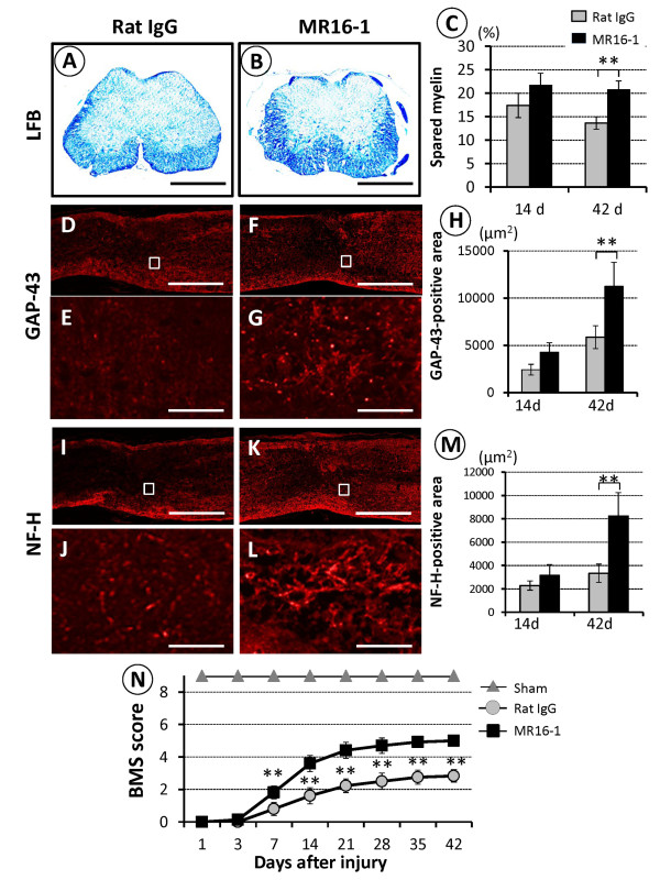
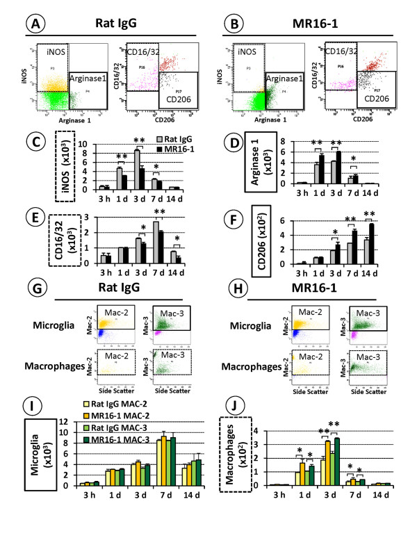
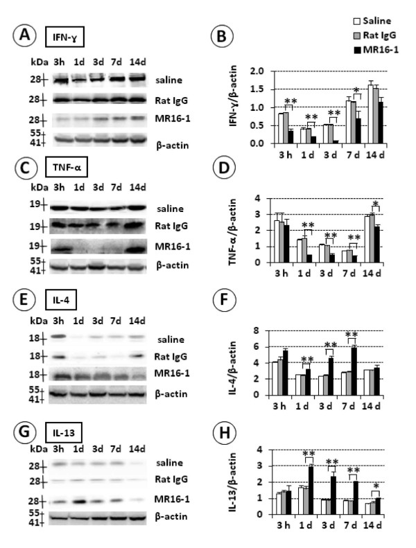
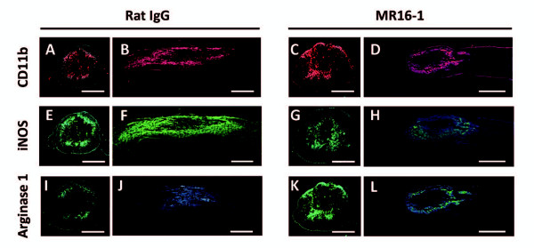
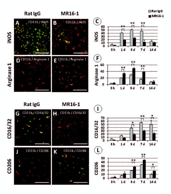
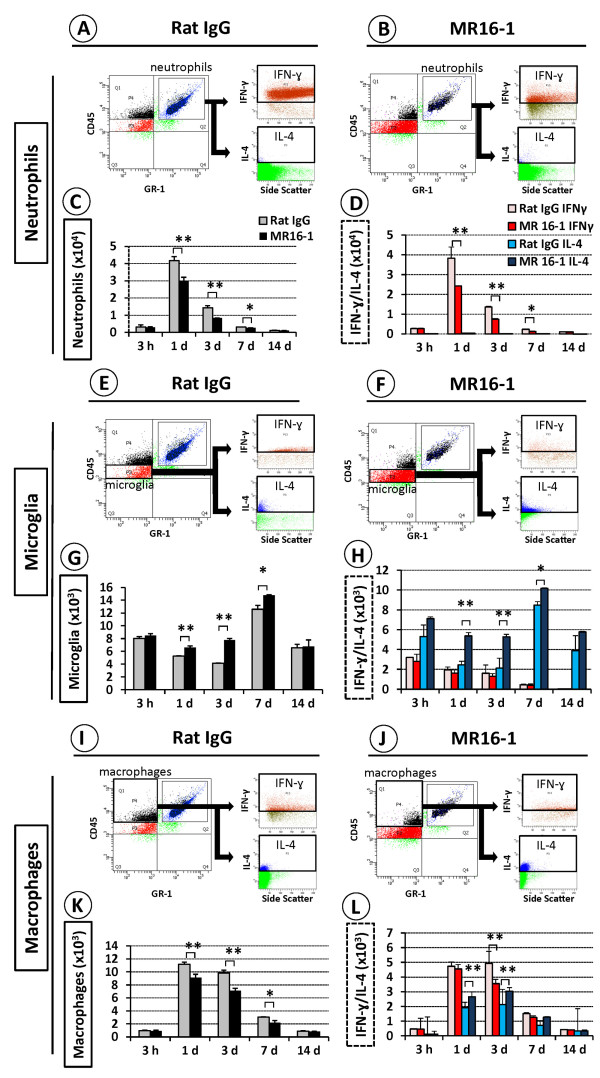
MR16-1 antibodies versus isotype control antibodies or saline alone were administered immediately after thoracic SCI in mice. SC tissue repair was compared between the two groups by Luxol fast blue (LFB) staining for myelination and immunoreactivity for the neuronal markers growth-associated protein (GAP)-43 and neurofilament heavy 200 kDa (NF-H) and for locomotor function. The expression of T helper (Th)1 cytokines (interferon (IFN)-γ and tumor necrosis factor-α) and Th2 cytokines (IL-4, IL-13) was determined by immunoblot analysis. The presence of M1 (inducible nitric oxide synthase (iNOS)-positive, CD16/32-positive) and M2 (arginase 1-positive, CD206-positive) macrophages was determined by immunohistology. Using flow cytometry, we also quantified IFN-γ and IL-4 levels in neutrophils, microglia, and macrophages, and Mac-2 (macrophage antigen-2) and Mac-3 in M2 macrophages and microglia.
LFB-positive spared myelin was increased in the MR16-1-treated group compared with the controls, and this increase correlated with enhanced positivity for GAP-43 or NF-H, and improved locomotor Basso Mouse Scale scores. Immunoblot analysis of the MR16-1-treated samples identified downregulation of Th1 and upregulation of Th2 cytokines. Whereas iNOS-positive, CD16/32-positive M1 macrophages were the predominant phenotype in the injured SC of non-treated control mice, MR16-1 treatment promoted arginase 1-positive, CD206-positive M2 macrophages, with preferential localization of these cells at the injury site. MR16-1 treatment suppressed the number of IFN-γ-positive neutrophils, and increased the number of microglia present and their positivity for IL-4. Among the arginase 1-positive M2 macrophages, MR16-1 treatment increased positivity for Mac-2 and Mac-3, suggestive of increased phagocytic behavior.
The results suggest that temporal blockade of IL-6 signaling after SCI abrogates damaging inflammatory activity and promotes functional recovery by promoting the formation of alternatively activated M2 macrophages.
最近在非神经元和神经元组织中的体内和体外研究表明,巨噬细胞激活的不同途径会导致细胞具有不同的特性。白细胞介素(IL)-6 触发经典激活的炎症巨噬细胞(M1 表型),而另一种激活的巨噬细胞(M2 表型)则具有抗炎作用。本研究的目的是阐明使用抗小鼠 IL-6R 单克隆抗体(MR16-1)暂时阻断 IL-6/IL-6 受体(IL-6R)结合对小鼠脊髓损伤(SCI)后急性期巨噬细胞激活和炎症反应的影响。
在 SCI 后立即向小鼠注射 MR16-1 抗体或同型对照抗体或生理盐水。通过对髓鞘的 Luxol 快速蓝(LFB)染色和对神经元标志物生长相关蛋白(GAP)-43 和神经丝重 200 kDa(NF-H)的免疫反应性以及运动功能,比较两组之间的 SC 组织修复情况。通过免疫印迹分析确定 Th1 细胞因子(干扰素(IFN)-γ和肿瘤坏死因子-α)和 Th2 细胞因子(IL-4、IL-13)的表达。通过免疫组织化学确定 M1(诱导型一氧化氮合酶(iNOS)阳性,CD16/32 阳性)和 M2(精氨酸酶 1 阳性,CD206 阳性)巨噬细胞的存在。通过流式细胞术,我们还定量了中性粒细胞、小胶质细胞和巨噬细胞中的 IFN-γ和 IL-4 水平,以及 M2 巨噬细胞和小胶质细胞中的 Mac-2(巨噬细胞抗原-2)和 Mac-3。
与对照组相比,MR16-1 治疗组的 LFB 阳性保留髓鞘增加,这种增加与 GAP-43 或 NF-H 的阳性增强以及运动 Basso 小鼠量表评分的改善相关。MR16-1 治疗样本的免疫印迹分析表明 Th1 下调和 Th2 上调。在未经治疗的对照组小鼠的损伤性 SCI 中,iNOS 阳性、CD16/32 阳性的 M1 巨噬细胞是主要表型,而 MR16-1 治疗促进了精氨酸酶 1 阳性、CD206 阳性的 M2 巨噬细胞的形成,这些细胞优先定位于损伤部位。MR16-1 治疗抑制了 IFN-γ 阳性中性粒细胞的数量,并增加了存在的小胶质细胞数量及其 IL-4 的阳性。在精氨酸酶 1 阳性的 M2 巨噬细胞中,MR16-1 治疗增加了 Mac-2 和 Mac-3 的阳性,提示吞噬作用增强。
这些结果表明,SCI 后 IL-6 信号的时间阻断通过促进另一种激活的 M2 巨噬细胞的形成来阻断损伤性炎症活动并促进功能恢复。