College of Pharmacy, Research Institute of Pharmaceutical Sciences, Kyungpook National University, Daegu, Korea.
J Thromb Haemost. 2012 Jun;10(6):1145-51. doi: 10.1111/j.1538-7836.2012.04671.x.
Recent results have indicated that polyphosphate, released by activated platelets, can function as a procoagulant to modulate the proteolytic activity of serine proteases of the blood clotting cascade.
To determine whether polyphosphate is involved in inducing signal transduction in cellular and animal models.
The effect of polyphosphate on human umbilical vein endothelial cells was examined by monitoring cell permeability, apoptosis and activation of NF-κB after treating cells with different concentrations of polyphosphate. Moreover, the expression of cell surface adhesion molecules (VCAM-1, ICAM-1 and E-selectin) and the adhesion of THP-1 cells to polyphosphate-treated cells were monitored using established methods. In the in vivo model, the pro-inflammatory effect of polyphosphate was assessed by monitoring vascular permeability and migration of leukocytes to the peritoneal cavity of mice injected with polyphosphate.
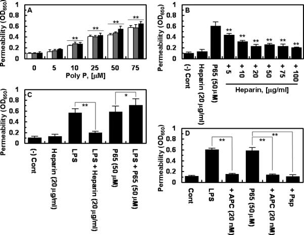
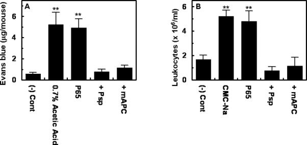
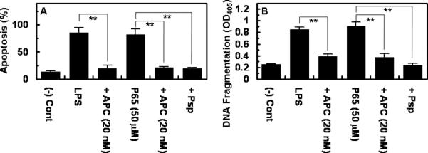
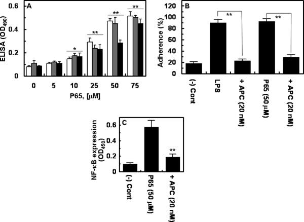
Polyphosphate, comprised of 45, 65 and 70 phosphate units, enhanced the barrier permeability and apoptosis in cultured endothelial cells and up-regulated the expression of cell adhesion molecules, thereby mediating the adhesion of THP-1 cells to polyphosphate-treated endothelial cells. These effects of polyphosphate were mediated through the activation of NF-κB and could not be recapitulated by another anionic polymer, heparin. Polyphosphate also increased the extravasation of the bovine serum albumin (BSA)-bound Evans blue dye and the migration of leukocytes to the mouse peritoneal cavity, which was prevented when activated protein C (APC) was intravenously (i.v.) injected 2 h before the challenge.
Polyphosphate, in addition to up-regulation of coagulation, can elicit potent pro-inflammatory responses through the activation of NF-κB, possibly contributing to the pro-inflammatory effect of activated platelets.
最近的研究结果表明,激活的血小板释放的多聚磷酸盐可以作为促凝剂,调节血液凝固级联中的丝氨酸蛋白酶的蛋白水解活性。
确定多聚磷酸盐是否参与细胞和动物模型中的信号转导。
通过监测不同浓度多聚磷酸盐处理后的细胞通透性、细胞凋亡和 NF-κB 的激活,研究多聚磷酸盐对人脐静脉内皮细胞的影响。此外,还通过建立的方法监测细胞表面黏附分子(VCAM-1、ICAM-1 和 E-选择素)的表达和 THP-1 细胞与多聚磷酸盐处理的细胞的黏附。在体内模型中,通过监测注射多聚磷酸盐的小鼠腹腔血管通透性和白细胞迁移来评估多聚磷酸盐的促炎作用。
由 45、65 和 70 个磷酸基组成的多聚磷酸盐增强了培养的内皮细胞的屏障通透性和细胞凋亡,并上调了细胞黏附分子的表达,从而介导了 THP-1 细胞与多聚磷酸盐处理的内皮细胞的黏附。多聚磷酸盐的这些作用是通过 NF-κB 的激活介导的,而另一种阴离子聚合物肝素则不能模拟。多聚磷酸盐还增加了牛血清白蛋白(BSA)结合 Evans 蓝染料的渗出量和白细胞向小鼠腹腔的迁移,而在挑战前 2 小时静脉注射激活蛋白 C(APC)可预防这种情况。
除了上调凝血作用外,多聚磷酸盐还可以通过激活 NF-κB 引发强烈的促炎反应,可能有助于激活血小板的促炎作用。