Center for Biomedical Engineering, The University of Texas Medical Branch, Galveston, TX, USA.
BMC Infect Dis. 2012 Feb 29;12:48. doi: 10.1186/1471-2334-12-48.
The development of safe topical microbicides that can preserve the integrity of cervicovaginal tract epithelial barrier is of great interest as this may minimize the potential for increased susceptibility to STI infections. High resolution imaging to assess epithelial integrity in a noninvasive manner could be a valuable tool for preclinical testing of candidate topical agents.
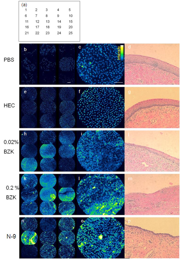
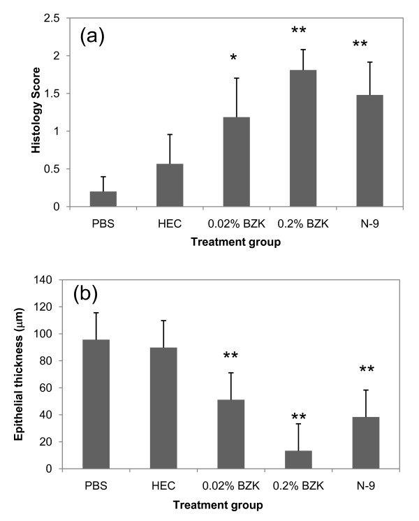
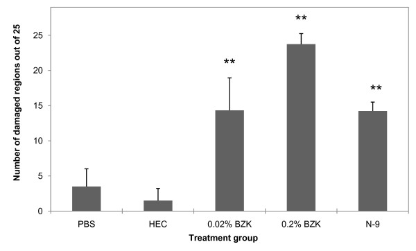
A quantitative approach using confocal fluorescence microendoscopy (CFM) for assessment of microbicide-induced injury to the vaginal epithelium was developed. Sheep were treated intravaginally with one of five agents in solution (PBS; 0.02% benzalkonium chloride (BZK); 0.2% BZK) or gel formulation (hydroxyethyl cellulose (HEC); Gynol II nonoxynol-9 gel (N-9)). After 24 hours the vaginal tract was removed, labeled with propidium iodide (PI), imaged, then fixed for histology. An automated image scoring algorithm was developed for quantitative assessment of injury and applied to the data set. Image-based findings were validated with histological visual gradings that describe degree of injury and measurement of epithelial thickness.
Distinct differences in PI staining were detected following BZK and N-9 treatment. Images from controls had uniformly distributed nuclei with defined borders, while those after BZK or N-9 showed heavily stained and disrupted nuclei, which increased in proportion to injury detected on histology. The confocal scoring system revealed statistically significant scores for each agent versus PBS controls with the exception of HEC and were consistent with histology scores of injury.
Confocal microendoscopy provides a sensitive, objective, and quantitative approach for non-invasive assessment of vaginal epithelial integrity and could serve as a tool for real-time safety evaluation of emerging intravaginal topical agents.
开发能够保持宫颈阴道上皮屏障完整性的安全局部杀微生物剂具有重要意义,因为这可能最大限度地降低增加对性传播感染易感性的风险。高分辨率成像技术可以非侵入性地评估上皮完整性,这可能是候选局部制剂临床前测试的有用工具。
我们开发了一种使用共聚焦荧光显微镜(CFM)评估阴道上皮杀微生物剂诱导损伤的定量方法。绵羊经阴道内给予五种溶液制剂(PBS;0.02%苯扎氯铵(BZK);0.2% BZK)或凝胶制剂(羟乙基纤维素(HEC);Gynol II 壬苯醇醚-9 凝胶(N-9))。24 小时后,取出阴道腔,用碘化丙啶(PI)标记,成像,然后固定用于组织学检查。开发了一种自动图像评分算法,用于对损伤进行定量评估,并应用于数据集。基于图像的发现与描述损伤程度和上皮厚度的组织学视觉分级进行了验证。
BZK 和 N-9 处理后,PI 染色有明显差异。对照图像中细胞核分布均匀,边界清晰,而 BZK 或 N-9 处理后的图像显示出染色强烈且紊乱的细胞核,其比例与组织学上检测到的损伤成正比。与 PBS 对照相比,CFM 评分系统显示出每个制剂的统计学显著评分,除了 HEC,并且与损伤的组织学评分一致。
共聚焦显微镜提供了一种敏感、客观和定量的方法,用于非侵入性评估阴道上皮完整性,并且可以作为实时评估新兴阴道内局部制剂安全性的工具。