Holstein Gay R, Friedrich Victor L, Martinelli Giorgio P, Ogorodnikov Dmitri, Yakushin Sergei B, Cohen Bernard
Department of Neurology, Mount Sinai School of Medicine New York, NY, USA.
Front Neurol. 2012 Feb 28;3:4. doi: 10.3389/fneur.2012.00004. eCollection 2012.
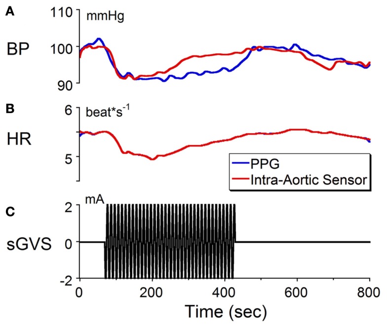
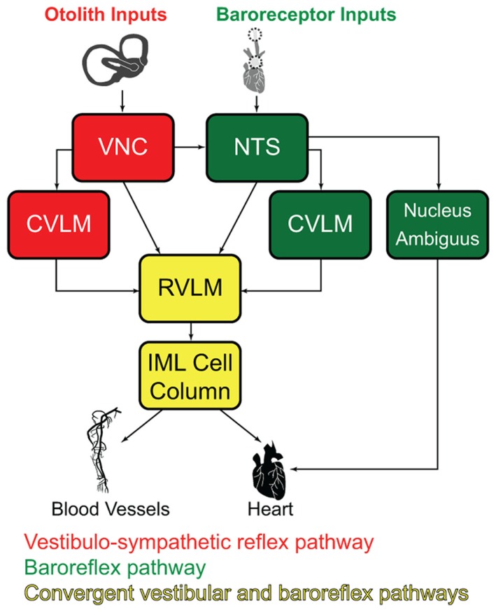
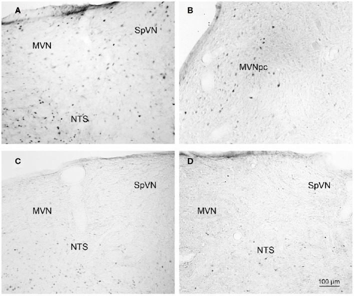
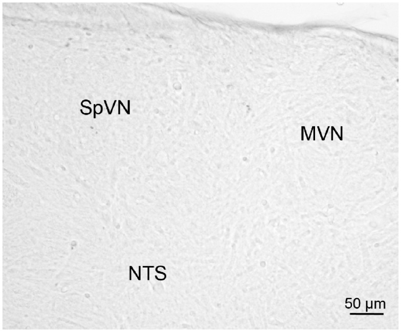
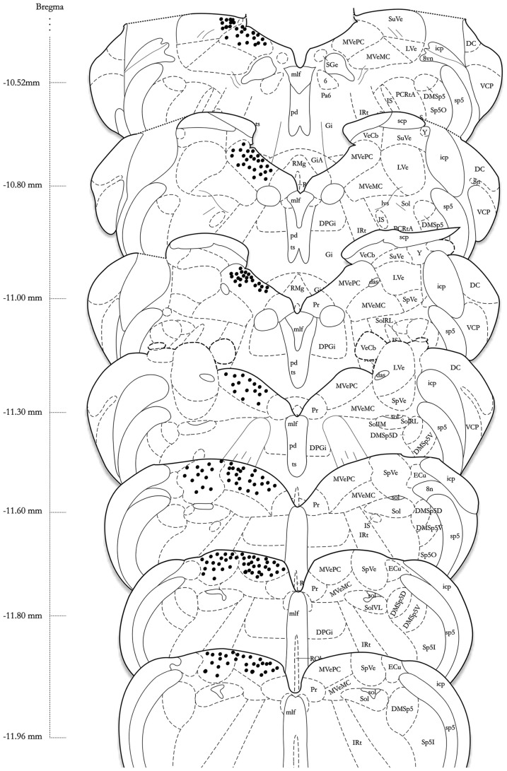
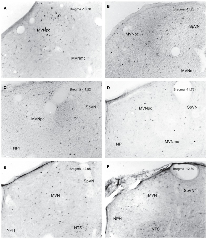
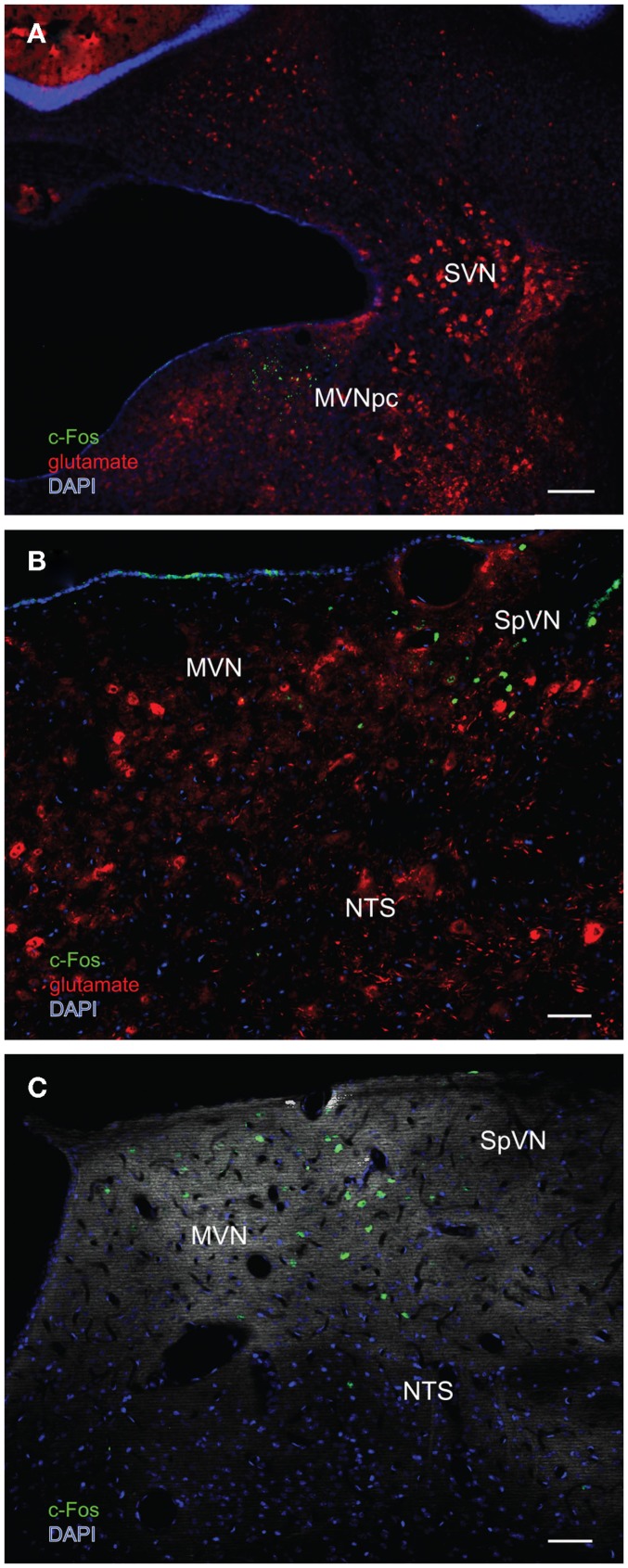
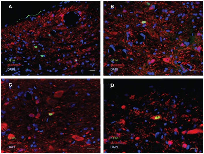
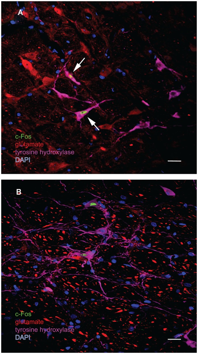
The vestibular system sends projections to brainstem autonomic nuclei that modulate heart rate and blood pressure in response to changes in head and body position with regard to gravity. Consistent with this, binaural sinusoidally modulated galvanic vestibular stimulation (sGVS) in humans causes vasoconstriction in the legs, while low frequency (0.02-0.04 Hz) sGVS causes a rapid drop in heart rate and blood pressure in anesthetized rats. We have hypothesized that these responses occur through activation of vestibulo-sympathetic pathways. In the present study, c-Fos protein expression was examined in neurons of the vestibular nuclei and rostral ventrolateral medullary region (RVLM) that were activated by low frequency sGVS. We found c-Fos-labeled neurons in the spinal, medial, and superior vestibular nuclei (SpVN, MVN, and SVN, respectively) and the parasolitary nucleus. The highest density of c-Fos-positive vestibular nuclear neurons was observed in MVN, where immunolabeled cells were present throughout the rostro-caudal extent of the nucleus. c-Fos expression was concentrated in the parvocellular region and largely absent from magnocellular MVN. c-Fos-labeled cells were scattered throughout caudal SpVN, and the immunostained neurons in SVN were restricted to a discrete wedge-shaped area immediately lateral to the IVth ventricle. Immunofluorescence localization of c-Fos and glutamate revealed that approximately one third of the c-Fos-labeled vestibular neurons showed intense glutamate-like immunofluorescence, far in excess of the stain reflecting the metabolic pool of cytoplasmic glutamate. In the RVLM, which receives a direct projection from the vestibular nuclei and sends efferents to preganglionic sympathetic neurons in the spinal cord, we observed an approximately threefold increase in c-Fos labeling in the sGVS-activated rats. We conclude that localization of c-Fos protein following sGVS is a reliable marker for sGVS-activated neurons of the vestibulo-sympathetic pathway.
前庭系统向脑干自主神经核发出投射,以响应头部和身体相对于重力的位置变化来调节心率和血压。与此一致的是,人类双耳正弦调制电流前庭刺激(sGVS)会导致腿部血管收缩,而低频(0.02 - 0.04Hz)sGVS会使麻醉大鼠的心率和血压迅速下降。我们推测这些反应是通过前庭 - 交感神经通路的激活而发生的。在本研究中,检测了由低频sGVS激活的前庭核和延髓头端腹外侧区(RVLM)神经元中的c - Fos蛋白表达。我们在脊髓、内侧和上前庭核(分别为SpVN、MVN和SVN)以及孤束旁核中发现了c - Fos标记的神经元。在MVN中观察到c - Fos阳性前庭核神经元的密度最高,在该核的整个 rostro - caudal范围内都存在免疫标记细胞。c - Fos表达集中在小细胞区域,大细胞MVN中基本没有。c - Fos标记的细胞散布在尾侧SpVN的整个区域,SVN中的免疫染色神经元局限于第四脑室紧邻外侧的一个离散楔形区域。c - Fos和谷氨酸的免疫荧光定位显示,约三分之一的c - Fos标记的前庭神经元呈现强烈的谷氨酸样免疫荧光,远远超过反映细胞质谷氨酸代谢池的染色。在接受前庭核直接投射并向脊髓节前交感神经元发出传出纤维的RVLM中,我们观察到sGVS激活的大鼠中c - Fos标记增加了约三倍。我们得出结论,sGVS后c - Fos蛋白的定位是前庭 - 交感神经通路中sGVS激活神经元的可靠标记。