Department of Neurology, Icahn School of Medicine at Mount Sinai , New York, NY , USA.
Department of Computer and Information Sciences, Brooklyn College of the City University of New York , Brooklyn, NY , USA.
Front Neurol. 2014 Apr 4;5:37. doi: 10.3389/fneur.2014.00037. eCollection 2014.
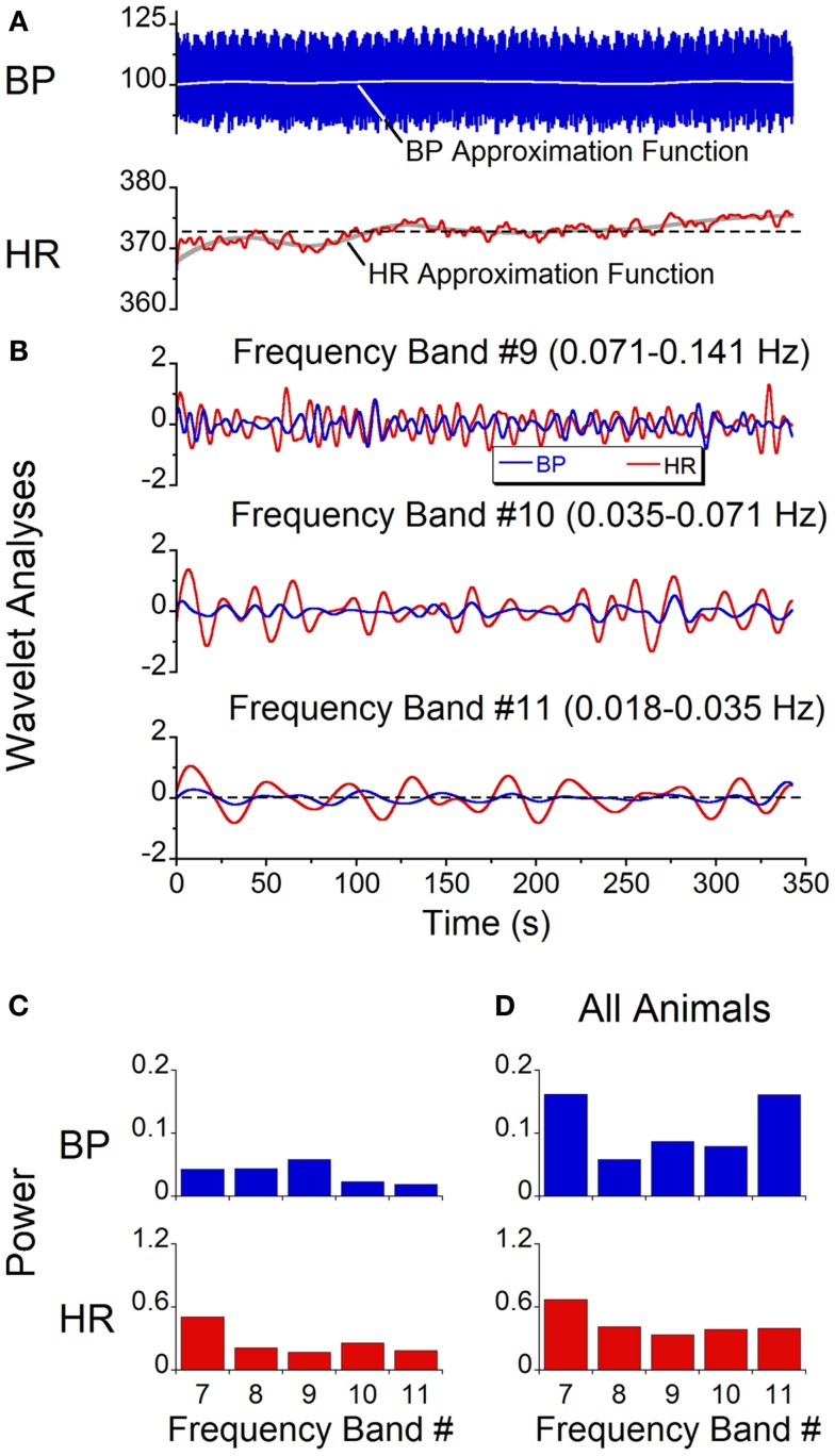
Sinusoidal galvanic vestibular stimulation (sGVS) induces oscillations in blood pressure (BP) and heart rate (HR), i.e., vasovagal oscillations, as well as transient decreases in BP and HR, i.e., vasovagal responses, in isoflurane-anesthetized rats. We determined the characteristics of the vasovagal oscillations, assessed their role in the generation of vasovagal responses, and determined whether they could be induced by monaural as well as by binaural sGVS and by oscillation in pitch. Wavelet analyses were used to determine the power distributions of the waveforms. Monaural and binaural sGVS and pitch generated vasovagal oscillations at the frequency and at twice the frequency of stimulation. Vasovagal oscillations and vasovagal responses were maximally induced at low stimulus frequencies (0.025-0.05 Hz). The oscillations were attenuated and the responses were rarely induced at higher stimulus frequencies. Vasovagal oscillations could occur without induction of vasovagal responses, but vasovagal responses were always associated with a vasovagal oscillation. We posit that the vasovagal oscillations originate in a low frequency band that, when appropriately activated by strong sympathetic stimulation, can generate vasovagal oscillations as a precursor for vasovagal responses and syncope. We further suggest that the activity responsible for the vasovagal oscillations arises in low frequency, otolith neurons with orientation vectors close to the vertical axis of the head. These neurons are likely to provide critical input to the vestibulo-sympathetic reflex to increase BP and HR upon changes in head position relative to gravity, and to contribute to the production of vasovagal oscillations and vasovagal responses and syncope when the baroreflex is inactivated.
窦房结电流前庭刺激 (sGVS) 会在异氟烷麻醉大鼠中引起血压 (BP) 和心率 (HR) 的振荡,即血管迷走神经振荡,以及 BP 和 HR 的短暂下降,即血管迷走神经反应。我们确定了血管迷走神经振荡的特征,评估了它们在血管迷走神经反应产生中的作用,并确定它们是否可以由单耳和双耳 sGVS 以及音高振荡引起。小波分析用于确定波形的功率分布。单耳和双耳 sGVS 以及音高刺激会在刺激频率和两倍刺激频率下产生血管迷走神经振荡。血管迷走神经振荡和血管迷走神经反应在低刺激频率 (0.025-0.05 Hz) 下最大程度地被诱导。当刺激频率较高时,振荡被减弱且反应很少被诱导。血管迷走神经振荡可以在不引起血管迷走神经反应的情况下发生,但血管迷走神经反应总是与血管迷走神经振荡相关联。我们假设血管迷走神经振荡起源于低频带,当受到强烈的交感神经刺激适当地激活时,它可以产生血管迷走神经振荡,作为血管迷走神经反应和晕厥的前兆。我们进一步提出,负责血管迷走神经振荡的活动起源于低频、耳石神经元,其取向向量接近头部的垂直轴。这些神经元可能为前庭交感反射提供关键输入,以在头部相对于重力的位置发生变化时增加 BP 和 HR,并在血压反射失活时有助于血管迷走神经振荡、血管迷走神经反应和晕厥的产生。