Laryngeal and Speech Section, National Institute of Neurological Disorders and Stroke, National Institutes of Health, Building 10, 5D-38, 10 Center Drive, MSC 1416, Bethesda, MD 20892-1416, USA.
Exp Brain Res. 2012 May;219(1):85-96. doi: 10.1007/s00221-012-3069-9. Epub 2012 Mar 23.
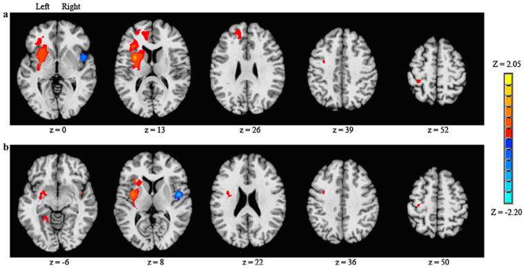
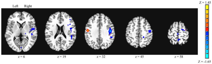
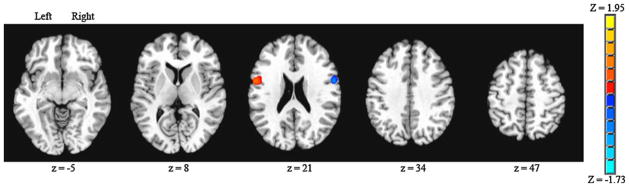
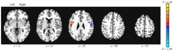
Functional neuroimaging has shown that multiple brain regions are active during volitional swallowing. Little is known, however, about which regions integrate motor execution and sensory feedback in the swallowing system. Although unilateral brain lesions in either hemisphere can produce swallowing deficits, some functional neuroimaging studies indicate that the left hemisphere has greater activation in certain sensory and motor-related swallowing regions. In this study, correlation coefficients were computed for five seed regions during volitional saliva swallowing to determine the functional relationships of these regions with the rest of the brain: the anterior and posterior insula, inferior frontal gyrus (BA44), primary sensory cortex (S1), and primary motor cortex (M1). A laterality index (LI) was derived that accounts for relative differences in total, positive connected voxels for the left/right hemisphere seeds. Clusters of significantly connected voxels were greater from the anterior and posterior insula than from the other three seed regions. Interactions of the insula with other brain regions were greater on the left than on the right during volitional swallowing. Group means showed laterality in the anterior insula (LI = 0.25) and the posterior insula (LI = 0.33). BA44 showed a lesser degree of difference in left versus right hemisphere interactions (LI = 0.12) while S1 did not show lateralization (LI = 0.02) and M1 showed some predominance of interactions in the right hemisphere (LI = -0.19). The greater connectivity from the left hemisphere insula to brain regions within and across hemispheres suggests that the insula is a primary integrative region for volitional swallowing in humans.
功能神经影像学研究表明,在自主吞咽过程中多个脑区活跃。然而,人们对哪些区域在吞咽系统中整合运动执行和感觉反馈知之甚少。尽管大脑单侧半球的病变都可能导致吞咽障碍,但一些功能神经影像学研究表明,左侧大脑半球在某些与感觉和运动相关的吞咽区域有更大的激活。在这项研究中,计算了五个种子区域在自主唾液吞咽期间的相关系数,以确定这些区域与大脑其余部分的功能关系:前岛叶和后岛叶、下额前回(BA44)、初级感觉皮层(S1)和初级运动皮层(M1)。得出了一个侧性指数(LI),该指数考虑了左/右半球种子的总阳性连接体素的相对差异。与其他三个种子区域相比,来自前岛叶和后岛叶的显著连接体素簇更大。在自主吞咽过程中,岛叶与大脑其他区域的相互作用在左侧大于右侧。组平均值显示前岛叶(LI=0.25)和后岛叶(LI=0.33)存在侧化。BA44 显示出左半球与右半球相互作用之间较小的差异(LI=0.12),而 S1 没有显示出侧化(LI=0.02),M1 显示出右半球相互作用的一些优势(LI=-0.19)。来自左侧岛叶的与大脑内和跨半球的区域的更大连通性表明,岛叶是人类自主吞咽的主要整合区域。