Department of Cardiology, West-German Heart Center Essen, Essen University Hospital, University Duisburg-Essen, Hufelandstrasse 55, Essen, Germany.
J Cardiovasc Magn Reson. 2012 Mar 27;14(1):21. doi: 10.1186/1532-429X-14-21.
Real-time cardiovascular magnetic resonance (rtCMR) is considered attractive for guiding TAVI. Owing to an unlimited scan plane orientation and an unsurpassed soft-tissue contrast with simultaneous device visualization, rtCMR is presumed to allow safe device navigation and to offer optimal orientation for precise axial positioning. We sought to evaluate the preclinical feasibility of rtCMR-guided transarterial aortic valve implatation (TAVI) using the nitinol-based Medtronic CoreValve bioprosthesis.
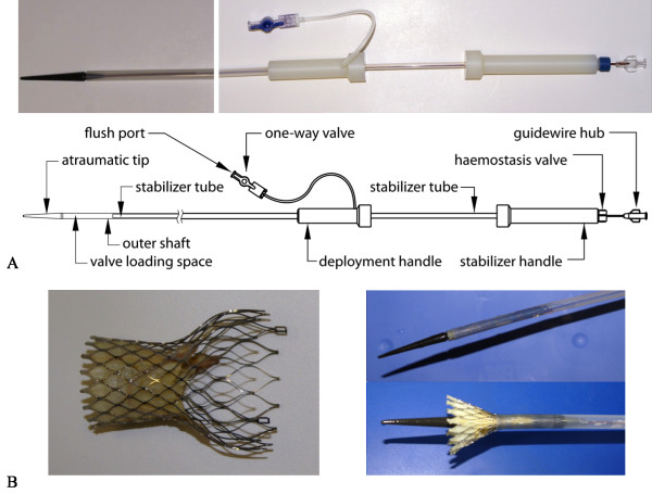
rtCMR-guided transfemoral (n = 2) and transsubclavian (n = 6) TAVI was performed in 8 swine using the original CoreValve prosthesis and a modified, CMR-compatible delivery catheter without ferromagnetic components.
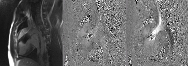
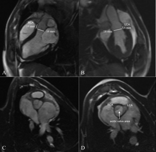
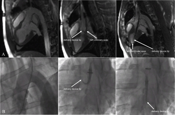
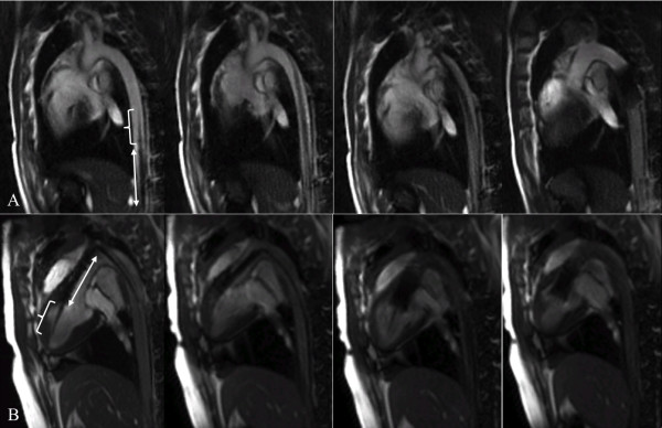
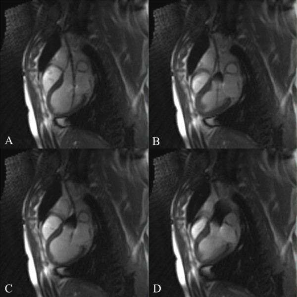
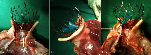
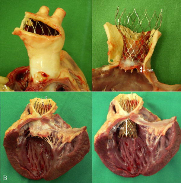
rtCMR using TrueFISP sequences provided reliable imaging guidance during TAVI, which was successful in 6 swine. One transfemoral attempt failed due to unsuccessful aortic arch passage and one pericardial tamponade with subsequent death occurred as a result of ventricular perforation by the device tip due to an operating error, this complication being detected without delay by rtCMR. rtCMR allowed for a detailed, simultaneous visualization of the delivery system with the mounted stent-valve and the surrounding anatomy, resulting in improved visualization during navigation through the vasculature, passage of the aortic valve, and during placement and deployment of the stent-valve. Post-interventional success could be confirmed using ECG-triggered time-resolved cine-TrueFISP and flow-sensitive phase-contrast sequences. Intended valve position was confirmed by ex-vivo histology.
Our study shows that rtCMR-guided TAVI using the commercial CoreValve prosthesis in conjunction with a modified delivery system is feasible in swine, allowing improved procedural guidance including immediate detection of complications and direct functional assessment with reduction of radiation and omission of contrast media.
实时心血管磁共振(rtCMR)被认为具有吸引力,可以用于指导 TAVI。由于扫描平面方向不受限制,并且具有无与伦比的软组织对比,可以同时显示设备,因此 rtCMR 被认为可以安全地进行器械导航,并为精确的轴向定位提供最佳方向。我们旨在评估使用基于镍钛诺的美敦力 CoreValve 生物假体进行 rtCMR 引导经皮主动脉瓣植入术(TAVI)的临床前可行性。
在 8 头猪中使用原始 CoreValve 假体和修改后的、无铁磁部件的 CMR 兼容输送导管,经股动脉(n = 2)和经锁骨下动脉(n = 6)进行 rtCMR 引导 TAVI。
使用 TrueFISP 序列进行的 rtCMR 为 TAVI 提供了可靠的成像指导,在 6 头猪中获得成功。一次经股动脉尝试因主动脉弓通过不成功而失败,一次心包填塞伴随后因器械尖端导致心室穿孔而死亡,这一并发症因操作失误而发生,该并发症被 rtCMR 及时发现。rtCMR 允许对输送系统与安装的支架瓣膜和周围解剖结构进行详细的、同时可视化,从而在通过血管导航、通过主动脉瓣以及在支架瓣膜的放置和部署过程中提高可视化效果。使用 ECG 触发的时间分辨 cine-TrueFISP 和流量敏感相位对比序列可以确认介入后的成功。预期的瓣膜位置通过离体组织学得到确认。
我们的研究表明,使用商业 CoreValve 假体和修改后的输送系统进行 rtCMR 引导的 TAVI 在猪中是可行的,允许进行改进的程序指导,包括并发症的即时检测和直接功能评估,同时减少辐射和省略对比剂。