Department of Biomedical Imaging and Department of Neuroscience, Genentech, Inc., South San Francisco, California, USA.
PLoS One. 2012;7(2):e31814. doi: 10.1371/journal.pone.0031814. Epub 2012 Feb 28.
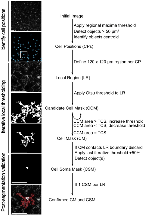
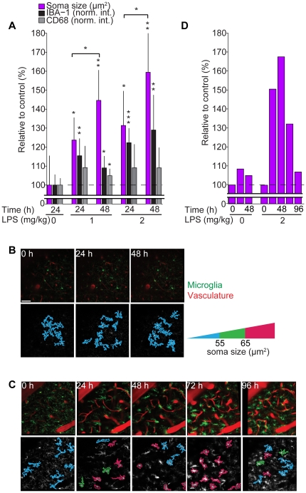
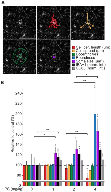
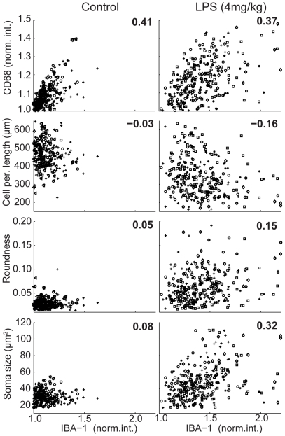
Microglia are specialized immune cells of the brain. Upon insult, microglia initiate a cascade of cellular responses including a characteristic change in cell morphology. To study the dynamics of microglia immune response in situ, we developed an automated image analysis method that enables the quantitative assessment of microglia activation state within tissue based solely on cell morphology. Per cell morphometric analysis of fluorescently labeled microglia is achieved through local iterative threshold segmentation, which reduces errors caused by signal-to-noise variation across large volumes. We demonstrate, utilizing systemic application of lipopolysaccharide as a model of immune challenge, that several morphological parameters, including cell perimeter length, cell roundness and soma size, quantitatively distinguish resting versus activated populations of microglia within tissue comparable to traditional immunohistochemistry methods. Furthermore, we provide proof-of-concept data that monitoring soma size enables the longitudinal assessment of microglia activation in the mouse neocortex imaged via 2-photon in vivo microscopy. The ability to quantify microglia activation automatically by shape alone allows unbiased and rapid analysis of both fixed and in vivo central nervous system tissue.
小胶质细胞是大脑中特化的免疫细胞。受到刺激后,小胶质细胞会引发一系列细胞反应,包括细胞形态的特征性变化。为了原位研究小胶质细胞免疫反应的动态性,我们开发了一种自动图像分析方法,该方法仅基于细胞形态即可对组织内小胶质细胞激活状态进行定量评估。通过局部迭代阈值分割对荧光标记的小胶质细胞进行逐细胞形态计量分析,可以减少大体积信号噪声变化引起的误差。我们利用脂多糖全身给药作为免疫挑战模型进行了验证,结果表明,包括细胞周长长度、细胞圆度和胞体大小在内的几个形态参数可定量区分组织内静息和激活的小胶质细胞群体,与传统免疫组织化学方法相当。此外,我们还提供了概念验证数据,表明监测胞体大小可通过双光子在体显微镜对活体小鼠新皮层进行成像,实现小胶质细胞激活的纵向评估。仅凭形状即可自动量化小胶质细胞激活的能力,允许对固定和活体中枢神经系统组织进行无偏和快速分析。