Cardiovascular Rehabilitation Unit, Department of Rehabilitation Sciences, Katholieke Universiteit Leuven, Tervuursevest 101, 3001 Heverlee, Belgium.
BMC Med Imaging. 2012 Apr 2;12:7. doi: 10.1186/1471-2342-12-7.
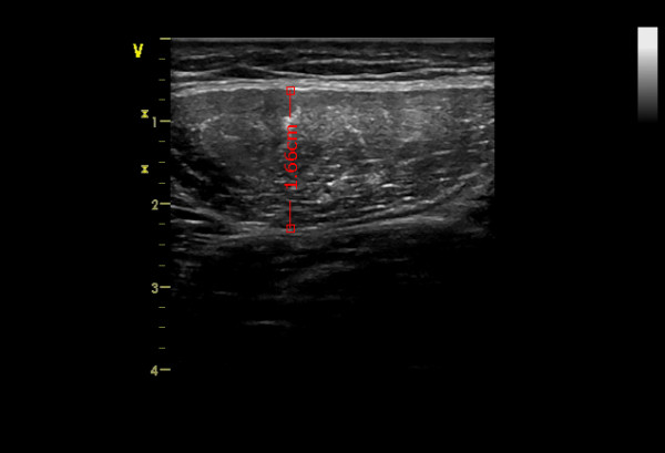
The increasing age of coronary artery disease (CAD) patients and the occurrence of sarcopenia in the elderly population accompanied by 'fear of moving' and hospitalization in these patients often results in a substantial loss of skeletal muscle mass and muscle strength. Cardiac rehabilitation can improve exercise tolerance and muscle strength in CAD patients but less data describe eventual morphological muscular changes possibly by more difficult access to imaging techniques. Therefore the aim of this study is to assess and quantify the reliability and validity of an easy applicable method, the ultrasound (US) technique, to measure the diameter of rectus femoris muscle in comparison to the muscle dimensions measured with CT scans.
45 older CAD patients without cardiac event during the last 9 months were included in this study. 25 patients were tested twice with ultrasound with a two day interval to assess test-retest reliability and 20 patients were tested twice (once with US and once with CT) on the same day to assess the validity of the US technique compared to CT as the gold standard. Isometric and isokinetic muscle testing was performed to test potential zero-order correlations between muscle diameter, muscle volume and muscle force.
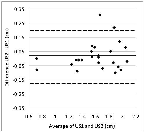
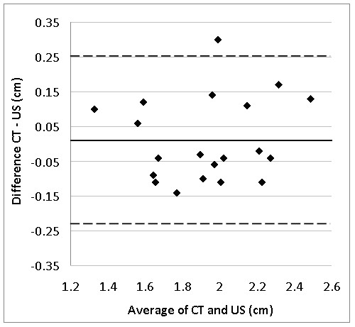
An intraclass correlation coefficient (ICC) of 0.97 ((95%CL: 0.92 - 0.99) was found for the test-retest reliability of US and the ICC computed between US and CT was 0.92 (95%CL: 0.81 - 0.97). The absolute difference between both techniques was 0.01 ± 0.12 cm (p = 0.66) resulting in a typical percentage error of 4.4%. Significant zero-order correlations were found between local muscle volume and muscle diameter assessed with CT (r = 0.67, p = 0.001) and assessed with US (r = 0.49, p < 0.05). Muscle strength parameters were also significantly correlated with muscle diameter assessed with both techniques (range r = 0.45-r = 0.61, p < 0.05).
Ultrasound imaging can be used as a valid and reliable measurement tool to assess the rectus femoris muscle diameter in older CAD patients.
冠心病(CAD)患者年龄的增加和老年人群中出现的肌肉减少症伴随着这些患者的“害怕活动”和住院,经常导致大量的骨骼肌质量和肌肉力量丧失。心脏康复可以提高 CAD 患者的运动耐量和肌肉力量,但关于最终的肌肉形态变化的数据较少,这可能是因为更难以获得成像技术。因此,本研究的目的是评估和量化一种简单应用的方法,即超声(US)技术,以测量股直肌直径,并与 CT 扫描测量的肌肉尺寸进行比较,以评估其可靠性和有效性。
本研究纳入了 45 名近 9 个月内无心脏事件的老年 CAD 患者。其中 25 名患者在两天的间隔内进行了两次超声检查,以评估测试-再测试的可靠性,20 名患者在同一天进行了两次检查(一次使用 US,一次使用 CT),以评估 US 技术与 CT 作为金标准的有效性。进行等长和等速肌肉测试,以测试肌肉直径、肌肉体积和肌肉力量之间潜在的零阶相关性。
US 的测试-再测试可靠性的组内相关系数(ICC)为 0.97(95%置信区间:0.92-0.99),US 与 CT 之间的 ICC 为 0.92(95%置信区间:0.81-0.97)。两种技术之间的绝对差异为 0.01±0.12cm(p=0.66),导致典型的百分比误差为 4.4%。在 CT 评估的局部肌肉体积(r=0.67,p=0.001)和 US 评估的局部肌肉体积(r=0.49,p<0.05)之间存在显著的零阶相关性。肌肉力量参数也与两种技术评估的肌肉直径呈显著相关(范围 r=0.45-r=0.61,p<0.05)。
超声成像可以作为一种有效的、可靠的测量工具,用于评估老年 CAD 患者的股直肌直径。