Department of Chemistry, m/c 111, University of Illinois at Chicago, Chicago, Illinois 60607-7061, USA.
Anal Chem. 2012 May 1;84(9):3945-51. doi: 10.1021/ac300557a. Epub 2012 Apr 16.
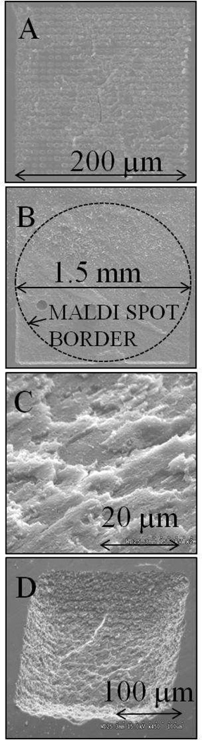
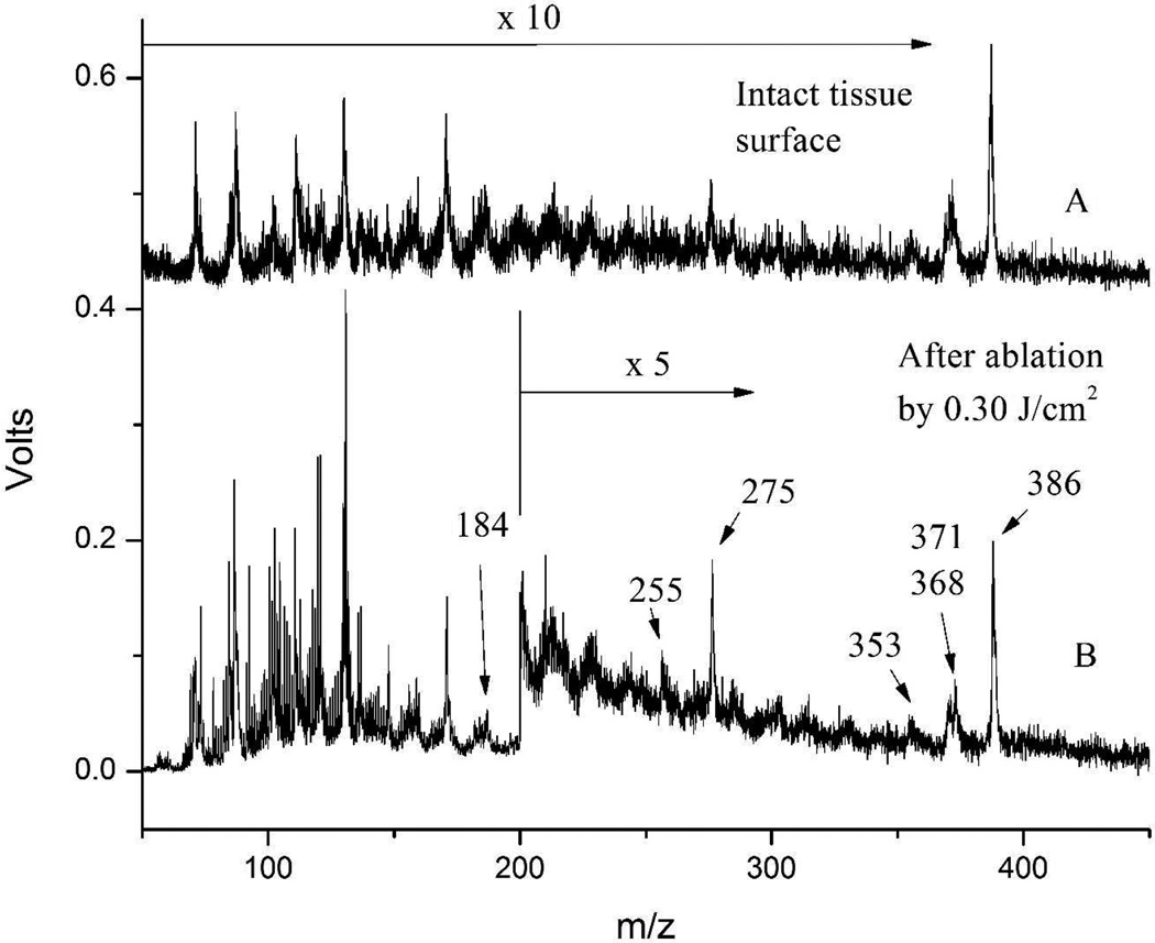
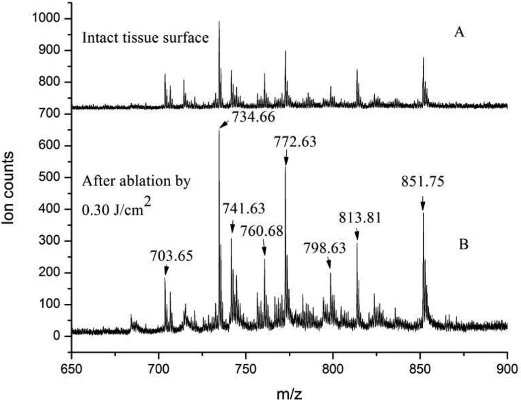
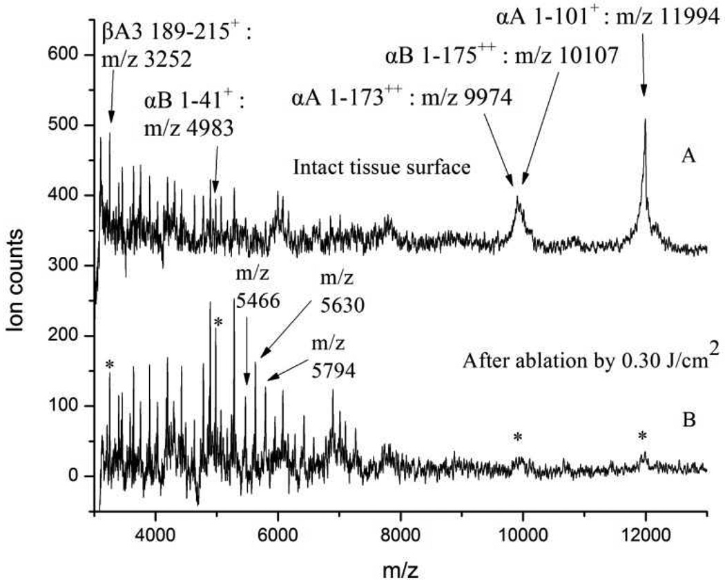
Experiments were performed to examine the feasibility of mass spectrometry (MS) depth profiling of animal tissue by ~75 fs, 800 nm laser pulses to expose underlying layers of tissue for subsequent MS analysis. Matrix assisted laser desorption ionization mass spectrometry (MALDI-MS) was used to analyze phospholipids and proteins from both intact bovine eye lens tissue and tissue ablated by ultrashort laser pulses. Laser desorption postionization mass spectrometry (LDPI-MS) with 10.5 eV single photon ionization was also used to analyze cholesterol and other small molecules in the tissue before and after laser ablation. Scanning electron microscopy was applied to examine the ablation patterns in the tissue and estimate the depth of the ablation craters. Ultrashort pulse laser ablation was found to be able to remove a layer of several tens of micrometers from the surface of eye lens tissue while leaving the underlying tissue relatively undamaged for subsequent MS analysis. MS analysis of cholesterol, phospholipids, peptides, and various unidentified species did not reveal any chemical damage caused by ultrashort pulse laser ablation for analytes smaller than ~6 kDa. However, a drop in intensity of larger protein ions was detected by MALDI-MS following laser ablation. An additional advantage was that ablated tissue displayed up to an order of magnitude higher signal intensities than intact tissue when subsequently analyzed by MS. These results support the use of ultrashort pulse laser ablation in combination with MS analysis to permit depth profiling of animal tissue.
实验旨在检验利用75fs、800nm 激光脉冲对动物组织进行质谱(MS)深度剖析的可行性,以暴露组织的下伏层,随后进行 MS 分析。基质辅助激光解吸电离质谱(MALDI-MS)用于分析完整牛眼晶状体组织和经超短激光脉冲烧蚀后的组织中的磷脂和蛋白质。还采用 10.5eV 单光子电离的激光解吸后电离质谱(LDPI-MS)分析激光烧蚀前后组织中的胆固醇和其他小分子。扫描电子显微镜用于检查组织中的烧蚀模式并估计烧蚀坑的深度。超短脉冲激光烧蚀能够从晶状体组织表面去除数十微米厚的一层,而使下伏组织相对不受损,以便随后进行 MS 分析。MS 分析胆固醇、磷脂、肽和各种未识别物种均未显示出小于6kDa 的分析物因超短脉冲激光烧蚀而产生的任何化学损伤。然而,在激光烧蚀后,MALDI-MS 检测到较大蛋白质离子的强度下降。另一个优点是,与完整组织相比,随后用 MS 分析时,烧蚀组织的信号强度高达一个数量级。这些结果支持将超短脉冲激光烧蚀与 MS 分析相结合,以实现动物组织的深度剖析。