Institute of Neuroscience and Medicine (INM-1, INM-2), Research Centre Jülich, 52425, Jülich, Germany.
Brain Struct Funct. 2013 Mar;218(2):511-26. doi: 10.1007/s00429-012-0411-8. Epub 2012 Apr 10.
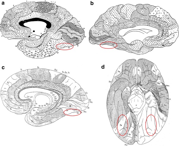
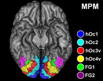
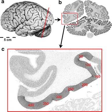
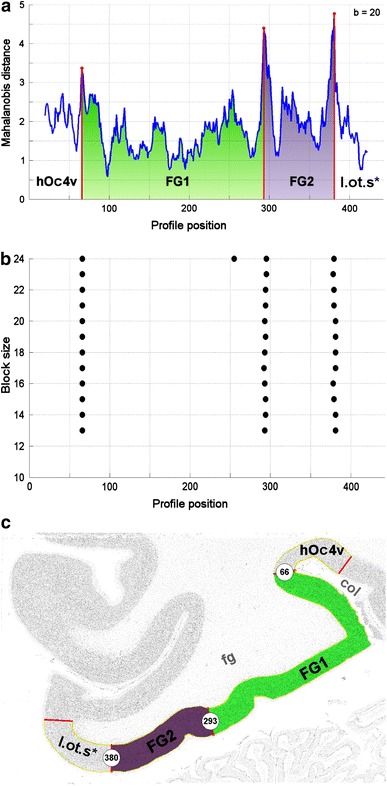
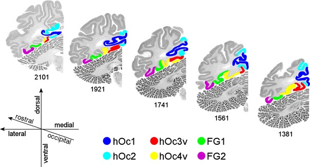
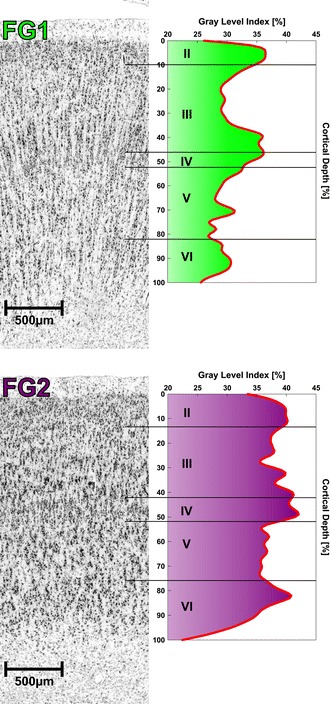
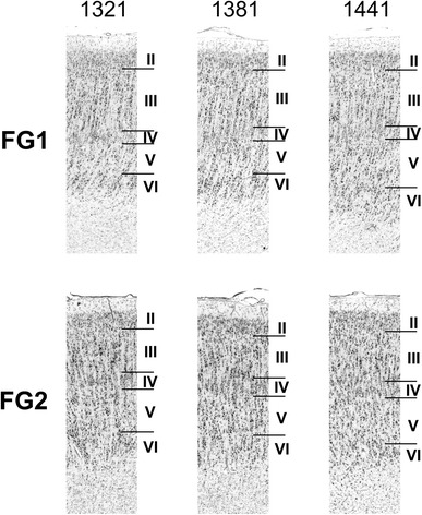
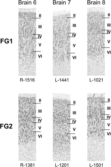
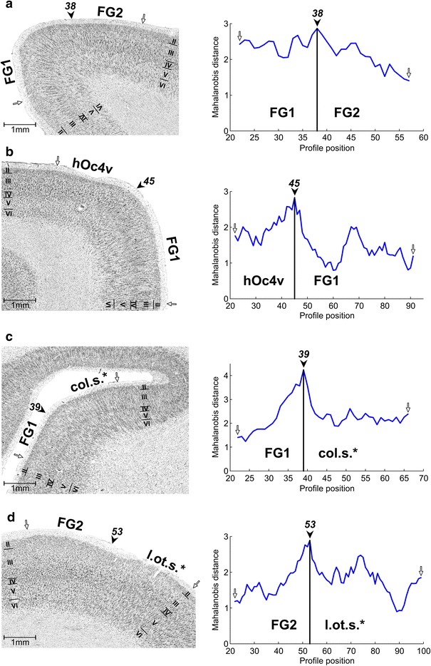
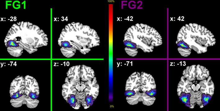
The human extrastriate visual cortex comprises numerous functionally defined areas, which are not identified in the widely used cytoarchitectonical map of Brodmann. The ventral part of the extrastriate cortex is particularly devoted to the identification of visual objects, faces and word forms. We analyzed the region immediately antero-lateral to hOc4v in serially sectioned (20 μm) and cell body-stained human brains using a quantitative observer-independent cytoarchitectonical approach to further identify the anatomical organization of the extrastriate cortex. Two novel cytoarchitectonical areas, FG1 and FG2, were identified on the posterior fusiform gyrus. The results of ten postmortem brains were then registered to their MRI volumes (acquired before histological processing), 3D reconstructed, and spatially normalized to the Montreal Neurological Institute reference brain. Finally, probabilistic maps were generated for each cytoarchitectonical area by superimposing the areas of the individual brains in the reference space. Comparison with recent functional imaging studies yielded that both areas are located within the object-related visual cortex. FG1 fills the gap between the retinotopically mapped area VO-1 and a posterior fusiform face patch. FG2 is probably the correlate of this face patch.
人类的外视皮层包含许多功能定义明确的区域,而这些区域在广泛使用的布罗德曼细胞构筑图谱中并未被识别出来。外视皮层的腹侧部分特别用于识别视觉物体、面孔和单词形式。我们使用定量的、观察者独立的细胞构筑学方法分析了在连续切片(20μm)和细胞体染色的人类大脑中,位于 hOc4v 前外侧的区域,以进一步确定外视皮层的解剖组织。在梭状回上发现了两个新的细胞构筑学区域 FG1 和 FG2。然后,将十个死后大脑的结果注册到他们的 MRI 体积(在组织学处理之前获得),进行 3D 重建,并以蒙特利尔神经学研究所参考大脑进行空间标准化。最后,通过在参考空间中叠加个体大脑的区域,为每个细胞构筑学区域生成概率图。与最近的功能成像研究进行比较,结果表明这两个区域都位于与物体相关的视觉皮层内。FG1 填补了视敏度映射区域 VO-1 和梭状回后部面孔区域之间的空白。FG2 可能是这个面孔区域的对应物。