Department of Biomedical Engineering, Calcium Signals Laboratory, Ross Building, Room 713, 720 Rutland Avenue, Baltimore, Maryland 21205-2196, USA.
Nat Commun. 2012 Apr 10;3:778. doi: 10.1038/ncomms1777.
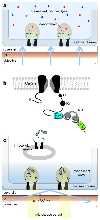
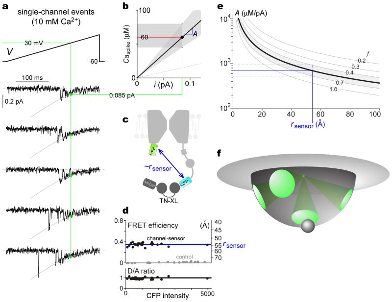
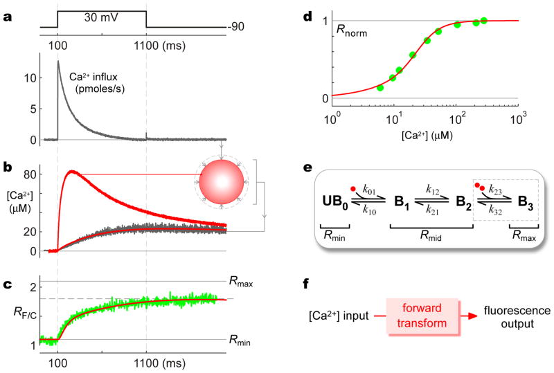
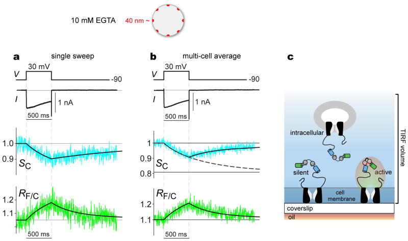
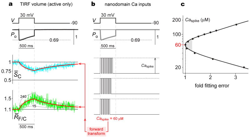
Coupling of excitation to secretion, contraction and transcription often relies on Ca(2+) computations within the nanodomain-a conceptual region extending tens of nanometers from the cytoplasmic mouth of Ca(2+) channels. Theory predicts that nanodomain Ca(2+) signals differ vastly from the slow submicromolar signals routinely observed in bulk cytoplasm. However, direct visualization of nanodomain Ca(2+) far exceeds optical resolution of spatially distributed Ca(2+) indicators. Here we couple an optical, genetically encoded Ca(2+) indicator (TN-XL) to the carboxy tail of Ca(V)2.2 Ca(2+) channels, enabling near-field imaging of the nanodomain. Under total internal reflection fluorescence microscopy, we detect Ca(2+) responses indicative of large-amplitude pulses. Single-channel electrophysiology reveals a corresponding Ca(2+) influx of only 0.085 pA, and fluorescence resonance energy transfer measurements estimate TN-XL distance to the cytoplasmic mouth at ~55 Å. Altogether, these findings raise the possibility that Ca(2+) exits the channel through the analogue of molecular portals, mirroring the crystallographic images of side windows in voltage-gated K channels.
兴奋与分泌、收缩和转录的偶联通常依赖于纳米域内的 Ca(2+)计算——这是一个从 Ca(2+)通道细胞质口延伸数十纳米的概念区域。理论预测,纳米域 Ca(2+)信号与通常在细胞质中观察到的缓慢亚微米级信号有很大的不同。然而,纳米域 Ca(2+)的直接可视化远远超出了空间分布 Ca(2+)指示剂的光学分辨率。在这里,我们将一种光学、基因编码的 Ca(2+)指示剂 (TN-XL) 与 Ca(V)2.2 Ca(2+)通道的羧基尾部偶联,从而能够对纳米域进行近场成像。在全内反射荧光显微镜下,我们检测到表明大振幅脉冲的 Ca(2+)响应。单通道电生理学显示相应的 Ca(2+)内流仅为 0.085 pA,荧光共振能量转移测量估计 TN-XL 距离细胞质口约为 55Å。总之,这些发现提出了一种可能性,即 Ca(2+)通过类似于分子门户的通道离开通道,反映了电压门控 K 通道侧窗的晶体学图像。