Medical Pharmacology and Physiology, University of Missouri, Columbia, MO 65212, USA.
Circ Res. 2012 May 11;110(10):1311-21. doi: 10.1161/CIRCRESAHA.111.262592. Epub 2012 Apr 5.
Electrical conduction through gap junction channels between endothelial cells of resistance vessels is integral to blood flow control. Small and intermediate-conductance Ca(2+)-activated K(+) channels (SK(Ca)/IK(Ca)) initiate electrical signals in endothelial cells, but it is unknown whether SK(Ca)/IK(Ca) activation alters signal transmission along the endothelium.
We tested the hypothesis that SK(Ca)/IK(Ca) activity regulates electrical conduction along the endothelium of resistance vessels.
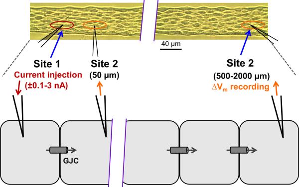
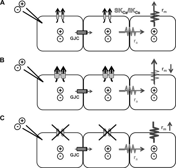
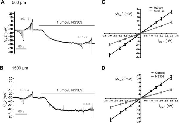
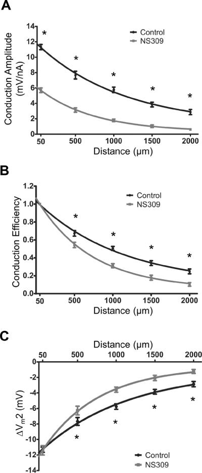
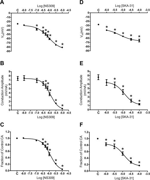
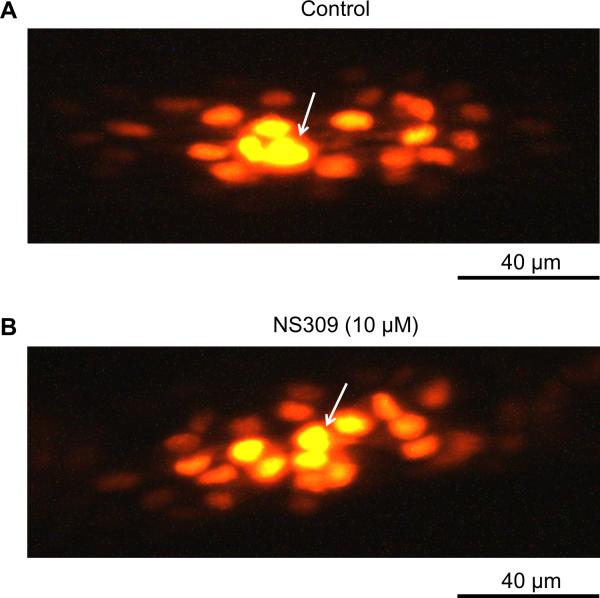
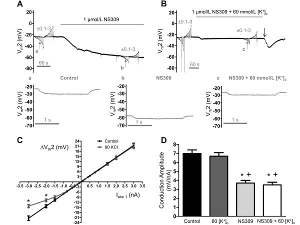
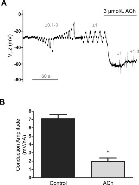
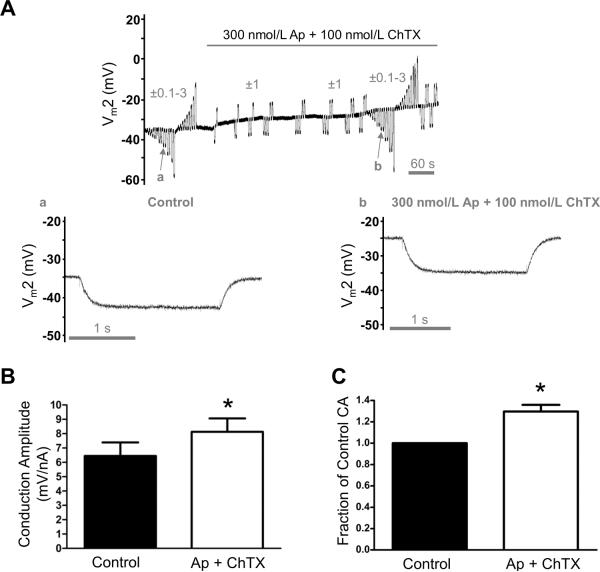
Freshly isolated endothelial cell tubes (60 μm wide; 1-3 mm long; cell length, ≈35 μm) from mouse skeletal muscle feed (superior epigastric) arteries were studied using dual intracellular microelectrodes. Current was injected (±0.1-3 nA) at site 1 while recording membrane potential (V(m)) at site 2 (separation distance=50-2000 μm). SK(Ca)/IK(Ca) activation (NS309, 1 μmol/L) reduced the change in V(m) along endothelial cell tubes by ≥50% and shortened the electrical length constant (λ) from 1380 to 850 μm (P<0.05) while intercellular dye transfer (propidium iodide) was maintained. Activating SK(Ca)/IK(Ca) with acetylcholine or SKA-31 also reduced electrical conduction. These effects of SK(Ca)/IK(Ca) activation persisted when hyperpolarization (>30 mV) was prevented with 60 mmol/L K(+). Conversely, blocking SK(Ca)/IK(Ca) (apamin+charybdotoxin) depolarized cells by ≈10 mV and enhanced electrical conduction (ie, changes in V(m)) by ≈30% (P<0.05).
These findings illustrate a novel role for SK(Ca)/IK(Ca) activity in tuning electrical conduction along the endothelium of resistance vessels by governing signal dissipation through changes in membrane resistance. Voltage-insensitive ion channels can thereby tune intercellular electrical signaling independent from gap junction channels.
电阻血管内皮细胞之间的缝隙连接通道的电传导对于血流控制至关重要。小电导和中等电导钙激活钾通道(SK(Ca)/IK(Ca))在血管内皮细胞中引发电信号,但尚不清楚 SK(Ca)/IK(Ca)的激活是否会改变沿内皮的信号传递。
我们检验了 SK(Ca)/IK(Ca)活性调节阻力血管内皮细胞电传导的假说。
使用双细胞内微电极研究了来自小鼠骨骼肌饲管(上腹)动脉的新鲜分离的内皮细胞管(60μm 宽;1-3mm 长;细胞长度约为 35μm)。在细胞 1 处注入电流(±0.1-3nA),同时在细胞 2 处记录膜电位(V(m))(分离距离=50-2000μm)。激活 SK(Ca)/IK(Ca)(NS309,1μmol/L)使内皮细胞管中 V(m)的变化减少了≥50%,并将电长度常数(λ)从 1380 缩短至 850μm(P<0.05),同时保持细胞间染料传递(碘化丙啶)。乙酰胆碱或 SKA-31 激活 SK(Ca)/IK(Ca)也会降低电传导。在使用 60mmol/L[K(+)](o)防止超极化(>30mV)时,SK(Ca)/IK(Ca)的激活仍保持这些作用。相反,阻断 SK(Ca)/IK(Ca)(apamin+charybdotoxin)使细胞去极化约 10mV,并增强电传导(即 V(m)的变化)约 30%(P<0.05)。
这些发现表明,SK(Ca)/IK(Ca)的活性通过改变膜电阻来控制信号耗散,从而在调节电阻血管内皮细胞的电传导中发挥了新的作用。因此,电压不敏感的离子通道可以独立于缝隙连接通道来调节细胞间的电信号。