Department of Medical Imaging of domestic animals and Orthopedics of small animals, Ghent University, Merelbeke, Belgium.
Acta Vet Scand. 2012 Apr 16;54(1):24. doi: 10.1186/1751-0147-54-24.
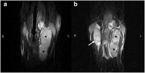
A 5-year-old castrated male Labrador Retriever was presented to a referring veterinarian for a swelling in the neck region. Based on the results of histopathology, a carotid body tumor, was diagnosed. The dog was referred to a medical imaging unit for further staging and follow up. This report describes the magnetic resonance (MR) and computed tomographic (CT) appearance of a carotid body tumor.
一只 5 岁已去势的雄性拉布拉多猎犬因颈部肿胀到转诊兽医处就诊。根据组织病理学结果,诊断为颈动脉体瘤。该犬被转诊到医学影像部门进行进一步分期和随访。本报告描述了颈动脉体瘤的磁共振(MR)和计算机断层(CT)表现。