Department of Musculoskeletal Biology, Institute of Ageing & Chronic Disease, University of Liverpool, Liverpool, Merseyside, United Kingdom.
PLoS One. 2012;7(4):e35299. doi: 10.1371/journal.pone.0035299. Epub 2012 Apr 11.
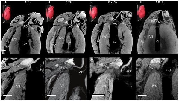
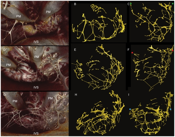
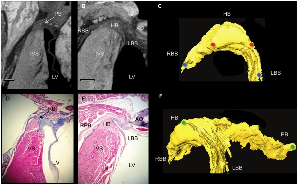
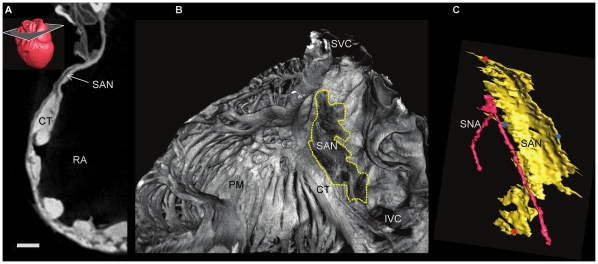
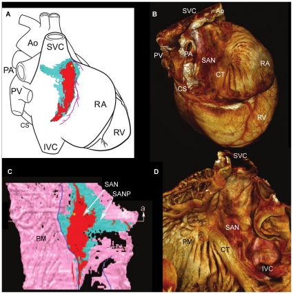
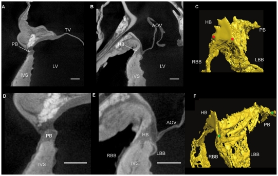
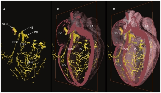
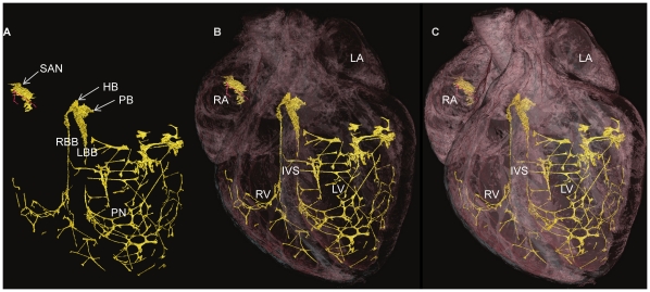
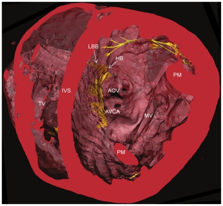
The general anatomy of the cardiac conduction system (CCS) has been known for 100 years, but its complex and irregular three-dimensional (3D) geometry is not so well understood. This is largely because the conducting tissue is not distinct from the surrounding tissue by dissection. The best descriptions of its anatomy come from studies based on serial sectioning of samples taken from the appropriate areas of the heart. Low X-ray attenuation has formerly ruled out micro-computed tomography (micro-CT) as a modality to resolve internal structures of soft tissue, but incorporation of iodine, which has a high molecular weight, into those tissues enhances the differential attenuation of X-rays and allows visualisation of fine detail in embryos and skeletal muscle. Here, with the use of a iodine based contrast agent (I(2)KI), we present contrast enhanced micro-CT images of cardiac tissue from rat and rabbit in which the three major subdivisions of the CCS can be differentiated from the surrounding contractile myocardium and visualised in 3D. Structures identified include the sinoatrial node (SAN) and the atrioventricular conduction axis: the penetrating bundle, His bundle, the bundle branches and the Purkinje network. Although the current findings are consistent with existing anatomical representations, the representations shown here offer superior resolution and are the first 3D representations of the CCS within a single intact mammalian heart.
心脏传导系统(CCS)的大体解剖结构已经为人所知 100 年了,但它复杂而不规则的三维(3D)几何形状却并不那么为人所理解。这在很大程度上是因为传导组织不能通过解剖与周围组织区分开来。对其解剖结构的最佳描述来自于基于从心脏适当区域取出的样本进行连续切片的研究。低 X 射线衰减以前排除了微计算机断层扫描(micro-CT)作为一种解决软组织内部结构的方法,但将具有高分子量的碘纳入这些组织中,增强了 X 射线的差异衰减,并允许在胚胎和骨骼肌中观察到精细的细节。在这里,我们使用碘基造影剂(I(2)KI),展示了来自大鼠和兔的心脏组织的增强对比度 micro-CT 图像,其中可以从周围的收缩心肌中区分出 CCS 的三个主要分支,并以 3D 形式可视化。确定的结构包括窦房结(SAN)和房室传导轴:穿透束、希氏束、束支和浦肯野纤维网。尽管目前的发现与现有的解剖学表示一致,但这里显示的表示提供了更高的分辨率,并且是单个完整哺乳动物心脏内 CCS 的第一个 3D 表示。