Department of Molecular Biology & Cytogenetics, Apollo Health City, Hyderabad, India.
Indian J Med Res. 2012 Mar;135(3):312-7.
BACKGROUND & OBJECTIVES: Fluorescence in situ hybridization (FISH) is increasingly being recognized as the most accurate and predictive test for HER 2/neu gene amplification and response to therapy in breast cancer. In the present study we investigated HER-2/neu gene amplification by FISH in breast carcinoma tissue specimens and compared the results with that of immunohistochemical (IHC) analysis.
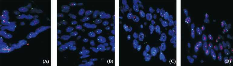
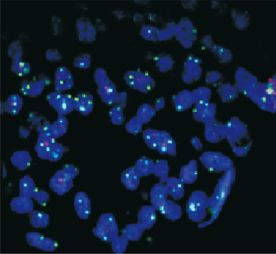
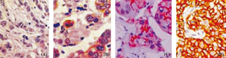
A total of 90 breast carcinoma tissue samples were used for immunohistochemical (IHC) and FISH analysis. IHC was performed by using mouse monoclonal antibody to the intracellular domain of HER-2/neu protein. Each slide was scored in a blinded fashion by two pathologists according to the manufacturer's recommended criteria. FISH analysis was performed on paraffin embedded breast tumour tissue sections. The polysomy for centromere 17 (Spec green signal) was read as green signals less than 4 as moderate polysomy, and more than 4 as highly polysomy.
Thirty of the 90 patients had negative results by IHC and FISH. Of the 28 patients with the score of 2+ by IHC, 20 were FISH positive for HER-2/neu gene amplification, three were FISH negative and five patients showed equivocal (1.8-2.2) results by FISH. These five cases were retested for IHC and FISH on different paraffin embedded tissue blocks, and all five were found positive for HER-2/neu gene amplification. Twenty five patients with the score of 3+ by IHC were FISH positive for HER-2/neu gene amplification (>2.2). Seven cases with the score of 3+ by IHC were FISH negative for HER-2/neu gene amplification (>2.2), and showed polysomy of chromosome number 17 high polysomy > 4.
INTERPRETATION & CONCLUSIONS: Our results indicated that HER-2/neu status by FISH should be performed in all cases of breast tumour with a 2+ score by IHC. Cases demonstrating a 3+ score by IHC may be subjected to FISH to rule out polysomy of chromosome 17 which could be falsely interpreted as HER-2/neu overexpression by IHC analysis. There is also a need for establishing a clinically validated cut-off value for HER-2/neu FISH amplification against IHC which may be further compared and calibrated.
荧光原位杂交(FISH)越来越被认为是乳腺癌中 HER2/neu 基因扩增和治疗反应最准确和有预测性的检测方法。本研究通过 FISH 检测乳腺癌组织标本中的 HER-2/neu 基因扩增,并将结果与免疫组织化学(IHC)分析进行比较。
共使用 90 例乳腺癌组织标本进行免疫组化(IHC)和 FISH 分析。IHC 采用针对 HER-2/neu 蛋白细胞内域的小鼠单克隆抗体进行。两位病理学家根据制造商推荐的标准对每张幻灯片进行盲法评分。FISH 分析在石蜡包埋的乳腺癌组织切片上进行。着丝粒 17 的三倍体(Spec 绿色信号)被读取为绿色信号小于 4 为中度三倍体,大于 4 为高度三倍体。
90 例患者中有 30 例 IHC 和 FISH 结果为阴性。28 例 IHC 评分 2+的患者中,20 例 FISH 检测到 HER-2/neu 基因扩增阳性,3 例 FISH 检测阴性,5 例患者 FISH 检测结果为(1.8-2.2)不确定。这 5 例患者在不同的石蜡包埋组织块上重新进行 IHC 和 FISH 检测,均检测到 HER-2/neu 基因扩增阳性。25 例 IHC 评分 3+的患者 FISH 检测到 HER-2/neu 基因扩增阳性(>2.2)。7 例 IHC 评分 3+的患者 FISH 检测到 HER-2/neu 基因扩增阴性(>2.2),并显示染色体 17 高倍数体(>4)。
我们的结果表明,所有 IHC 评分 2+的乳腺癌病例均应进行 FISH 检测 HER-2/neu 状态。IHC 评分 3+的病例可能需要进行 FISH 检测,以排除可能被 IHC 分析错误地解释为 HER-2/neu 过表达的染色体 17 三倍体。还需要建立针对 IHC 的 HER-2/neu FISH 扩增的临床验证截断值,然后可以进一步进行比较和校准。