Eberhardsteiner Lukas, Hellmich Christian, Scheiner Stefan
a Institute for Transportation Science, Research Center for Road Engineering, Vienna University of Technology , Vienna , Austria.
Comput Methods Biomech Biomed Engin. 2014;17(1):48-63. doi: 10.1080/10255842.2012.670227. Epub 2012 May 8.
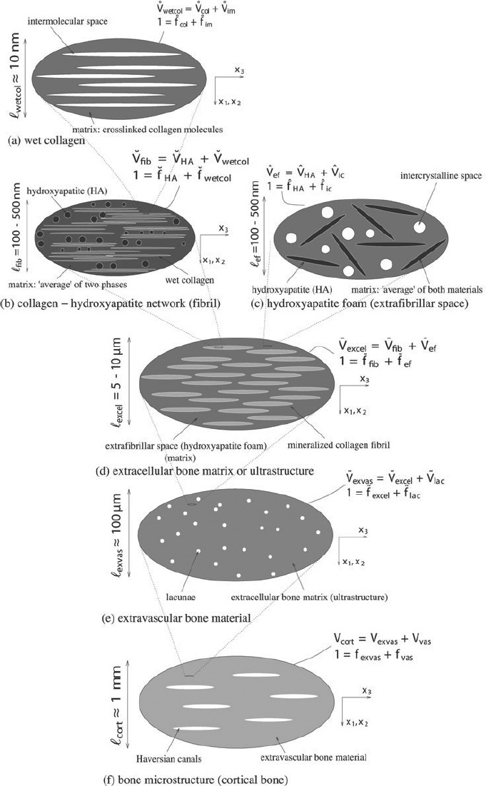
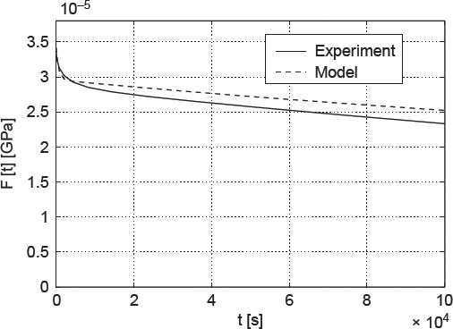
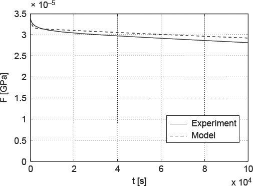
Extracellular bone material can be characterised as a nanocomposite where, in a liquid environment, nanometre-sized hydroxyapatite crystals precipitate within as well as between long fibre-like collagen fibrils (with diameters in the 100 nm range), as evidenced from neutron diffraction and transmission electron microscopy. Accordingly, these crystals are referred to as 'interfibrillar mineral' and 'extrafibrillar mineral', respectively. From a topological viewpoint, it is probable that the mineralisations start on the surfaces of the collagen fibrils ('mineral-encrusted fibrils'), from where the crystals grow both into the fibril and into the extrafibrillar space. Since the mineral concentration depends on the pore spaces within the fibrils and between the fibrils (there is more space between them), the majority of the crystals (but clearly not all of them) typically lie in the extrafibrillar space. There, larger crystal agglomerations or clusters, spanning tens to hundreds of nanometers, develop in the course of mineralisation, and the micromechanics community has identified the pivotal role, which this extrafibrillar mineral plays for tissue elasticity. In such extrafibrillar crystal agglomerates, single crystals are stuck together, their surfaces being covered with very thin water layers. Recently, the latter have caught our interest regarding strength properties (Fritsch et al. 2009 J Theor Biol. 260(2): 230-252) - we have identified these water layers as weak interfaces in the extrafibrillar mineral of bone. Rate-independent gliding effects of crystals along the aforementioned interfaces, once an elastic threshold is surpassed, can be related to overall elastoplastic material behaviour of the hierarchical material 'bone'. Extending this idea, the present paper is devoted to viscous gliding along these interfaces, expressing itself, at the macroscale, in the well-known experimentally evidenced phenomenon of bone viscoelasticity. In this context, a multiscale homogenisation scheme is extended to viscoelasticity, mineral-cluster-specific creep parameters are identified from three-point bending tests on hydrated bone samples, and the model is validated by statistically and physically independent experiments on partially dried samples. We expect this model to be relevant when it comes to prediction of time-dependent phenomena, e.g. in the context of bone remodelling.
细胞外骨材料可被视为一种纳米复合材料,在液体环境中,纳米级的羟基磷灰石晶体在长纤维状胶原纤维内部以及之间沉淀(纤维直径在100纳米范围内),中子衍射和透射电子显微镜观察证实了这一点。因此,这些晶体分别被称为“纤维间矿物质”和“纤维外矿物质”。从拓扑学角度来看,矿化作用很可能始于胶原纤维表面(“矿化包被纤维”),晶体从这里向纤维内部和纤维外空间生长。由于矿物质浓度取决于纤维内部和纤维之间的孔隙空间(它们之间的空间更大),大多数晶体(但显然不是全部)通常位于纤维外空间。在那里,在矿化过程中会形成直径达数十至数百纳米的更大晶体团聚体或簇,微观力学领域已经确定了这种纤维外矿物质对组织弹性所起的关键作用。在这种纤维外晶体团聚体中,单晶粘在一起,其表面覆盖着非常薄的水层。最近,后者在强度特性方面引起了我们的兴趣(弗里奇等人,《理论生物学杂志》2009年第260卷第2期,第230 - 252页)——我们已将这些水层确定为骨纤维外矿物质中的薄弱界面。一旦超过弹性阈值,晶体沿上述界面的与速率无关的滑动效应可能与“骨”这种分级材料的整体弹塑性材料行为有关。拓展这一概念,本文致力于研究沿这些界面的粘性滑动,这种滑动在宏观层面表现为骨粘弹性这一众所周知的实验证实现象。在此背景下,一种多尺度均匀化方案被扩展至粘弹性,通过对水合骨样本的三点弯曲试验确定了特定矿物簇的蠕变参数,并通过对部分干燥样本进行的统计和物理独立实验对该模型进行了验证。我们预计,在预测与时间相关的现象时,例如在骨重塑的背景下,该模型将具有相关性。