• 文献检索
  • 文档翻译
  • 深度研究
  • 学术资讯
  • Suppr Zotero 插件Zotero 插件
  • 邀请有礼
  • 套餐&价格
  • 历史记录
应用&插件
Suppr Zotero 插件Zotero 插件浏览器插件Mac 客户端Windows 客户端微信小程序
定价
高级版会员购买积分包购买API积分包
服务
文献检索文档翻译深度研究API 文档MCP 服务
关于我们
关于 Suppr公司介绍联系我们用户协议隐私条款
关注我们

Suppr 超能文献

核心技术专利:CN118964589B侵权必究
粤ICP备2023148730 号-1Suppr @ 2026

文献检索

告别复杂PubMed语法,用中文像聊天一样搜索,搜遍4000万医学文献。AI智能推荐,让科研检索更轻松。

立即免费搜索

文件翻译

保留排版,准确专业,支持PDF/Word/PPT等文件格式,支持 12+语言互译。

免费翻译文档

深度研究

AI帮你快速写综述,25分钟生成高质量综述,智能提取关键信息,辅助科研写作。

立即免费体验

巴西利什曼原虫分离株磷脂酰丝氨酸暴露与皮肤利什曼病的弥漫性和寄生虫感染力有关。

Exposure of phosphatidylserine on Leishmania amazonensis isolates is associated with diffuse cutaneous leishmaniasis and parasite infectivity.

机构信息

Centro de Pesquisas Gonçalo Moniz/FIOCRUZ-BA, Salvador, Brasil.

出版信息

PLoS One. 2012;7(5):e36595. doi: 10.1371/journal.pone.0036595. Epub 2012 May 4.

DOI:10.1371/journal.pone.0036595
PMID:22574191
原文链接:https://pmc.ncbi.nlm.nih.gov/articles/PMC3344919/
Abstract

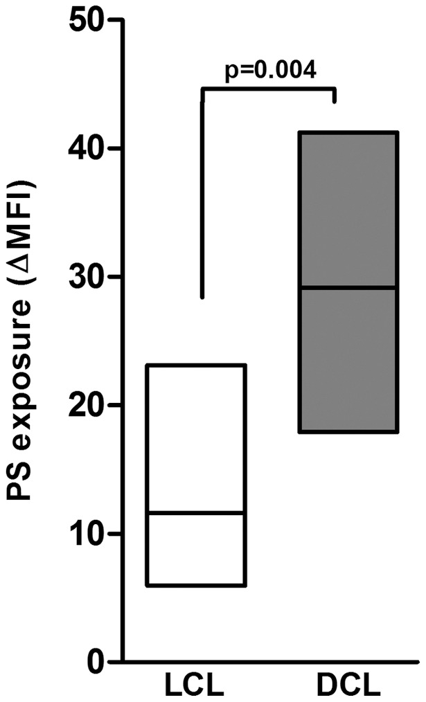
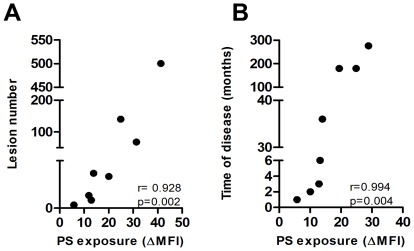
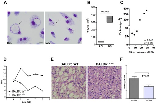
Diffuse cutaneous leishmaniasis (DCL) is a rare clinical manifestation of leishmaniasis, characterized by an inefficient parasite-specific cellular response and heavily parasitized macrophages. In Brazil, Leishmania (Leishmania) amazonensis is the main species involved in DCL cases. In the experimental model, recognition of phosphatidylserine (PS) molecules exposed on the surface of amastigotes forms of L. amazonensis inhibits the inflammatory response of infected macrophages as a strategy to evade the host immune surveillance. In this study, we examined whether PS exposure on L. amazonensis isolates from DCL patients operated as a parasite pathogenic factor and as a putative suppression mechanism of immune response during the infection. Peritoneal macrophages from F1 mice (BALB/c×C57BL/6) were infected with different L. amazonensis isolates from patients with localized cutaneous leishmaniasis (LCL) or DCL. DCL isolates showed higher PS exposure than their counterparts from LCL patients. In addition, PS exposure was positively correlated with clinical parameters of the human infection (number of lesions and time of disease) and with characteristics of the experimental infection (macrophage infection and anti-inflammatory cytokine induction). Furthermore, parasites isolated from DCL patients displayed an increased area in parasitophorous vacuoles (PV) when compared to those isolated from LCL patients. Thus, this study shows for the first time that a parasite factor (exposed PS) might be associated with parasite survival/persistence in macrophages and lesion exacerbation during the course of DCL, providing new insights regarding pathogenic mechanism in this rare chronic disease.

摘要

弥漫性皮肤利什曼病(DCL)是利什曼病的一种罕见临床表现,其特征是寄生虫特异性细胞应答效率低下,并且大量寄生的巨噬细胞。在巴西,Leishmania(Leishmania) amazonensis 是与 DCL 病例相关的主要物种。在实验模型中,识别暴露在 L. amazonensis 无鞭毛体表面的磷脂酰丝氨酸(PS)分子会抑制感染巨噬细胞的炎症反应,这是一种逃避宿主免疫监视的策略。在这项研究中,我们研究了 DCL 患者的 L. amazonensis 分离株表面 PS 的暴露是否作为寄生虫致病因素以及作为感染期间免疫应答抑制的潜在机制。F1 小鼠(BALB/c×C57BL/6)的腹腔巨噬细胞被来自局限性皮肤利什曼病(LCL)或 DCL 患者的不同 L. amazonensis 分离株感染。DCL 分离株的 PS 暴露水平高于来自 LCL 患者的分离株。此外,PS 暴露与人类感染的临床参数(病变数量和疾病时间)以及实验感染的特征(巨噬细胞感染和抗炎细胞因子诱导)呈正相关。此外,与来自 LCL 患者的分离株相比,来自 DCL 患者的寄生虫在吞噬空泡(PV)中的面积增加。因此,这项研究首次表明,寄生虫因子(暴露的 PS)可能与巨噬细胞中的寄生虫存活/持续存在以及 DCL 期间病变恶化有关,为这种罕见慢性疾病的发病机制提供了新的见解。

https://cdn.ncbi.nlm.nih.gov/pmc/blobs/1905/3344919/706d31459cbf/pone.0036595.g005.jpg
https://cdn.ncbi.nlm.nih.gov/pmc/blobs/1905/3344919/09a3c3a860c6/pone.0036595.g001.jpg
https://cdn.ncbi.nlm.nih.gov/pmc/blobs/1905/3344919/df9bba830878/pone.0036595.g002.jpg
https://cdn.ncbi.nlm.nih.gov/pmc/blobs/1905/3344919/e499ade5a07a/pone.0036595.g003.jpg
https://cdn.ncbi.nlm.nih.gov/pmc/blobs/1905/3344919/31df7fa5c571/pone.0036595.g004.jpg
https://cdn.ncbi.nlm.nih.gov/pmc/blobs/1905/3344919/706d31459cbf/pone.0036595.g005.jpg

相似文献

1
Exposure of phosphatidylserine on Leishmania amazonensis isolates is associated with diffuse cutaneous leishmaniasis and parasite infectivity.巴西利什曼原虫分离株磷脂酰丝氨酸暴露与皮肤利什曼病的弥漫性和寄生虫感染力有关。
PLoS One. 2012;7(5):e36595. doi: 10.1371/journal.pone.0036595. Epub 2012 May 4.
2
Differential Regulation of l-Arginine Metabolism through Arginase 1 during Infection with Leishmania mexicana Isolates Obtained from Patients with Localized and Diffuse Cutaneous Leishmaniasis.通过 Arg1ase1 对 l-精氨酸代谢的差异调控在来源于局限性和皮肤弥散性利什曼病患者的 Leishmania mexicana 分离株感染中的作用。
Infect Immun. 2020 Jun 22;88(7). doi: 10.1128/IAI.00963-19.
3
Host and parasite responses in human diffuse cutaneous leishmaniasis caused by L. amazonensis.由 L. amazonensis 引起的人类弥漫性皮肤利什曼病中的宿主和寄生虫反应。
PLoS Negl Trop Dis. 2019 Mar 7;13(3):e0007152. doi: 10.1371/journal.pntd.0007152. eCollection 2019 Mar.
4
PD-L1 May Mediate T-Cell Exhaustion in a Case of Early Diffuse Leishmaniasis Caused by (L.) .PD-L1 可能介导了由 (L.)引起的早期弥漫性利什曼病中的 T 细胞耗竭。
Front Immunol. 2018 May 11;9:1021. doi: 10.3389/fimmu.2018.01021. eCollection 2018.
5
Resistance of Leishmania (Leishmania) amazonensis and Leishmania (Viannia) braziliensis to nitric oxide correlates with disease severity in Tegumentary Leishmaniasis.亚马逊利什曼原虫(利什曼原虫属)和巴西利什曼原虫(维扬尼利什曼原虫属)对一氧化氮的抗性与皮肤利什曼病的疾病严重程度相关。
BMC Infect Dis. 2007 Feb 22;7:7. doi: 10.1186/1471-2334-7-7.
6
CD8 cells of patients with diffuse cutaneous leishmaniasis display functional exhaustion: the latter is reversed, in vitro, by TLR2 agonists.弥漫性皮肤利什曼病患者的 CD8 细胞表现出功能衰竭:后者可被 TLR2 激动剂体外逆转。
PLoS Negl Trop Dis. 2010 Nov 2;4(11):e871. doi: 10.1371/journal.pntd.0000871.
7
Experimental model of chronic osteomyelitis caused by Leishmania (L) amazonensis.亚马逊利什曼原虫(L)引起的慢性骨髓炎实验模型。
Acta Trop. 2006 May;98(2):125-9. doi: 10.1016/j.actatropica.2006.02.007. Epub 2006 May 11.
8
Isolation of intact Leishmania amazonensis large parasitophorous vacuoles from infected macrophages by density gradient fractionation.通过密度梯度分级分离从感染的巨噬细胞中分离完整的亚马逊利什曼原虫大滋养体空泡。
Exp Parasitol. 2020 Nov;218:107989. doi: 10.1016/j.exppara.2020.107989. Epub 2020 Sep 15.
9
Regulation of Leishmania (L.) amazonensis protein expression by host T cell dependent responses: differential expression of oligopeptidase B, tryparedoxin peroxidase and HSP70 isoforms in amastigotes isolated from BALB/c and BALB/c nude mice.宿主T细胞依赖性反应对亚马逊利什曼原虫蛋白质表达的调控:从BALB/c和BALB/c裸鼠分离的无鞭毛体中寡肽酶B、锥虫硫氧还蛋白过氧化物酶和HSP70亚型的差异表达
PLoS Negl Trop Dis. 2015 Feb 18;9(2):e0003411. doi: 10.1371/journal.pntd.0003411. eCollection 2015 Feb.
10
In vitro and in vivo miltefosine susceptibility of a Leishmania amazonensis isolate from a patient with diffuse cutaneous leishmaniasis.来自一名弥漫性皮肤利什曼病患者的亚马逊利什曼原虫分离株的体外和体内米替福新敏感性
PLoS Negl Trop Dis. 2014 Jul 17;8(7):e2999. doi: 10.1371/journal.pntd.0002999. eCollection 2014 Jul.

引用本文的文献

1
Isolation and characterisation of Leishmania (Leishmania) infantum from cutaneous leishmaniasis patients in northeast Brazil.从巴西东北部皮肤利什曼病患者中分离和鉴定利什曼原虫(利什曼原虫)。
Mem Inst Oswaldo Cruz. 2024 Jul 8;119:e240026. doi: 10.1590/0074-02760240026. eCollection 2024.
2
Genetic characterization and description of Leishmania (Leishmania) ellisi sp. nov.: a new human-infecting species from the USA.遗传特征分析与 Leishmania (Leishmania) ellisi sp. nov. 的描述:一种来自美国的新的人类感染物种。
Parasitol Res. 2023 Dec 15;123(1):52. doi: 10.1007/s00436-023-08034-8.
3
VAMP3 and VAMP8 Regulate the Development and Functionality of Parasitophorous Vacuoles Housing Leishmania amazonensis.

本文引用的文献

1
Cooperation between apoptotic and viable metacyclics enhances the pathogenesis of Leishmaniasis.凋亡性与存活的循环前鞭毛体之间的合作增强了利什曼病的发病机制。
PLoS One. 2009 May 29;4(5):e5733. doi: 10.1371/journal.pone.0005733.
2
Control of parasitophorous vacuole expansion by LYST/Beige restricts the intracellular growth of Leishmania amazonensis.LYST/米色蛋白对寄生泡扩张的控制限制了亚马逊利什曼原虫的细胞内生长。
PLoS Pathog. 2008 Oct;4(10):e1000179. doi: 10.1371/journal.ppat.1000179. Epub 2008 Oct 17.
3
Role of natural killer cells in modulating dendritic cell responses to Leishmania amazonensis infection.
VAMP3 和 VAMP8 调节寄生空泡的发育和功能,这些空泡容纳 Leishmania amazonensis。
Infect Immun. 2022 Mar 17;90(3):e0018321. doi: 10.1128/IAI.00183-21. Epub 2022 Feb 7.
4
-Induced Dendritic Cell Migration and Its Potential Contribution to Parasite Dissemination.诱导树突状细胞迁移及其对寄生虫播散的潜在作用。
Microorganisms. 2021 Jun 11;9(6):1268. doi: 10.3390/microorganisms9061268.
5
Mapping Alterations Induced by Long-Term Axenic Cultivation of Promastigotes With a Multiplatform Metabolomic Fingerprint Approach.采用多平台代谢组指纹图谱方法对无培养基长期培养的前鞭毛体诱导的变化进行映射。
Front Cell Infect Microbiol. 2019 Dec 4;9:403. doi: 10.3389/fcimb.2019.00403. eCollection 2019.
6
Draft Genome Sequences of () , () , and () , Potential Etiological Agents of Diffuse Cutaneous Leishmaniasis.弥漫性皮肤利什曼病的潜在病原体——()、()和()的基因组序列草图
Microbiol Resour Announc. 2019 May 16;8(20):e00269-19. doi: 10.1128/MRA.00269-19.
7
CD4 T Cell-Dependent Macrophage Activation Modulates Sustained PS Exposure on Intracellular Amastigotes of .CD4 T 细胞依赖性巨噬细胞激活调节细胞内无鞭毛体持续暴露于 PS 上。
Front Cell Infect Microbiol. 2019 Apr 12;9:105. doi: 10.3389/fcimb.2019.00105. eCollection 2019.
8
Systems Approach Reveals Nuclear Factor Erythroid 2-Related Factor 2/Protein Kinase R Crosstalk in Human Cutaneous Leishmaniasis.系统方法揭示人类皮肤利什曼病中核因子红细胞2相关因子2/蛋白激酶R的相互作用
Front Immunol. 2017 Sep 15;8:1127. doi: 10.3389/fimmu.2017.01127. eCollection 2017.
9
Modeling Immune Response to Species Indicates Adenosine As an Important Inhibitor of Th-Cell Activation.建模对 物种的免疫反应表明腺苷是 T 细胞活化的重要抑制剂。
Front Cell Infect Microbiol. 2017 Jul 20;7:309. doi: 10.3389/fcimb.2017.00309. eCollection 2017.
10
Cysteine Peptidase B Regulates Leishmania mexicana Virulence through the Modulation of GP63 Expression.半胱氨酸肽酶B通过调节GP63表达来调控墨西哥利什曼原虫的毒力。
PLoS Pathog. 2016 May 18;12(5):e1005658. doi: 10.1371/journal.ppat.1005658. eCollection 2016 May.
自然杀伤细胞在调节树突状细胞对亚马逊利什曼原虫感染反应中的作用。
Infect Immun. 2008 Nov;76(11):5100-9. doi: 10.1128/IAI.00438-08. Epub 2008 Sep 15.
4
In vivo imaging reveals an essential role for neutrophils in leishmaniasis transmitted by sand flies.体内成像揭示了中性粒细胞在白蛉传播的利什曼病中的重要作用。
Science. 2008 Aug 15;321(5891):970-4. doi: 10.1126/science.1159194.
5
Down-regulation of dendritic cell signaling pathways by Leishmania amazonensis amastigotes.亚马逊利什曼原虫无鞭毛体对树突状细胞信号通路的下调作用
Mol Immunol. 2008 Jul;45(12):3371-82. doi: 10.1016/j.molimm.2008.04.018. Epub 2008 Jun 5.
6
Vaccinia virus uses macropinocytosis and apoptotic mimicry to enter host cells.痘苗病毒利用巨胞饮作用和凋亡模拟来进入宿主细胞。
Science. 2008 Apr 25;320(5875):531-5. doi: 10.1126/science.1155164.
7
Role of interleukin-1beta in activating the CD11c(high) CD45RB- dendritic cell subset and priming Leishmania amazonensis-specific CD4+ T cells in vitro and in vivo.白细胞介素-1β在体外和体内激活CD11c(高)CD45RB-树突状细胞亚群以及启动亚马逊利什曼原虫特异性CD4+ T细胞中的作用。
Infect Immun. 2007 Oct;75(10):5018-26. doi: 10.1128/IAI.00499-07. Epub 2007 Aug 6.
8
Resistance of Leishmania (Leishmania) amazonensis and Leishmania (Viannia) braziliensis to nitric oxide correlates with disease severity in Tegumentary Leishmaniasis.亚马逊利什曼原虫(利什曼原虫属)和巴西利什曼原虫(维扬尼利什曼原虫属)对一氧化氮的抗性与皮肤利什曼病的疾病严重程度相关。
BMC Infect Dis. 2007 Feb 22;7:7. doi: 10.1186/1471-2334-7-7.
9
Endocytic recycling in yeast is regulated by putative phospholipid translocases and the Ypt31p/32p-Rcy1p pathway.酵母中的内吞循环由假定的磷脂转位酶和Ypt31p/32p-Rcy1p途径调控。
Mol Biol Cell. 2007 Jan;18(1):295-312. doi: 10.1091/mbc.e06-05-0461. Epub 2006 Nov 8.
10
Molecular diagnosis of leishmaniasis: current status and future applications.利什曼病的分子诊断:现状与未来应用
J Clin Microbiol. 2007 Jan;45(1):21-5. doi: 10.1128/JCM.02029-06. Epub 2006 Nov 8.