Department of Radiation Oncology, University of Florida, 2033 Mowry Road, Cancer Genetic Research Complex, Room 485E, Gainesville, FL 32610, USA.
BMC Cancer. 2012 May 28;12:198. doi: 10.1186/1471-2407-12-198.
The c-Met receptor tyrosine kinase is aberrantly activated in many solid tumors. In a prior study we showed that prostate cancer PC-3 cells exhibit constitutively activated c-Met without exogenous hepatocyte growth factor (HGF); however whether this characteristic is due to an endogenous HGF/c-Met autocrine loop remains controversial. In the current study we examined the response of PC-3 cells to an anti-HGF neutralizing antibody or a small molecule Met kinase inhibitor (BMS-777607).
Cell scattering was tested by monitoring cell morphology after HGF stimulation. Cell migration was examined by both "wound-healing" and transwell assasy and invasion was detected by Matrigel-coated transwell assay. Proliferation, survival and anoikis were determined by MTT, colony formation and trypan blue exclusion assay, respectively. Gene and protein expression were assessed by real-time PCR and Western blot, respectively.
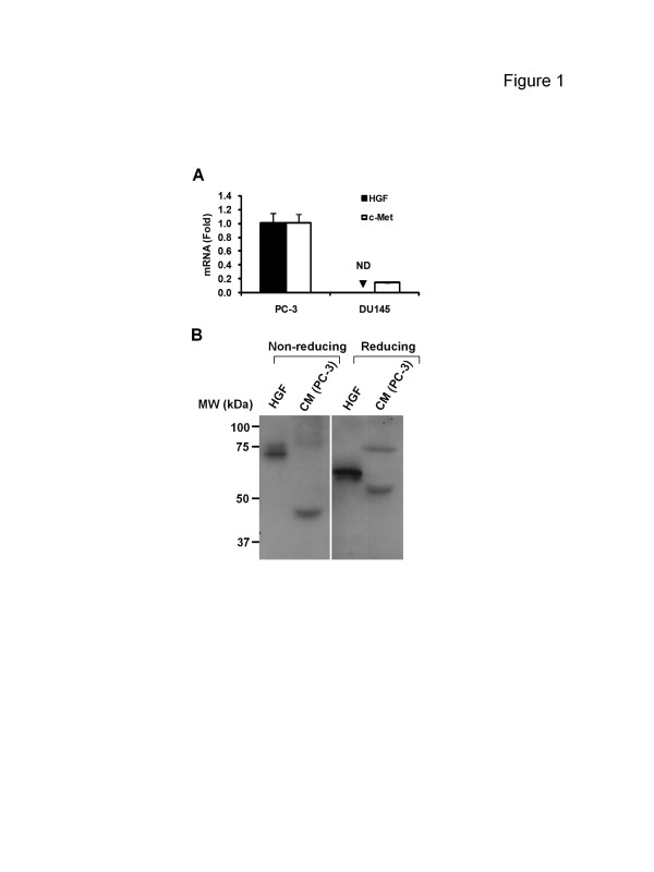
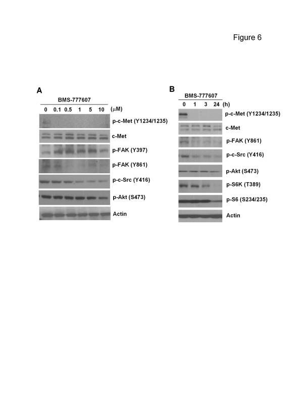
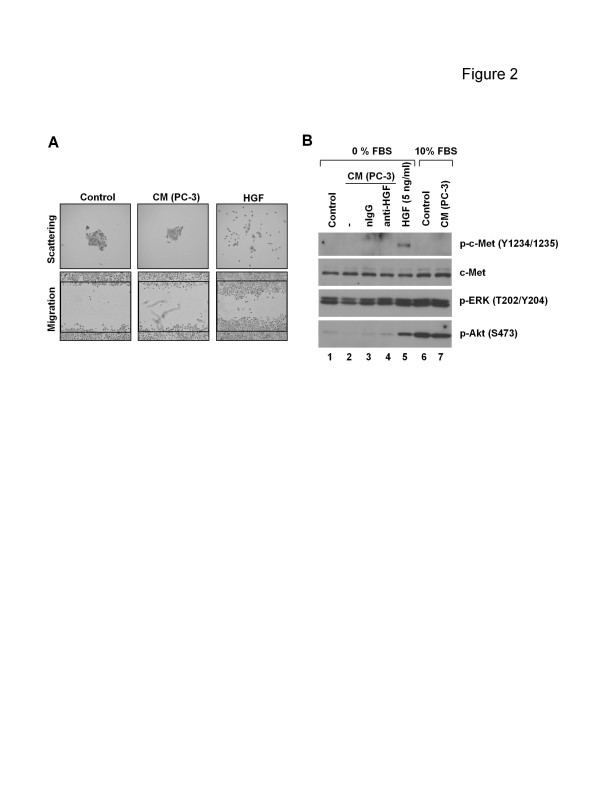
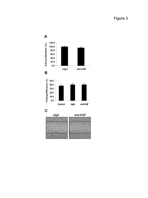
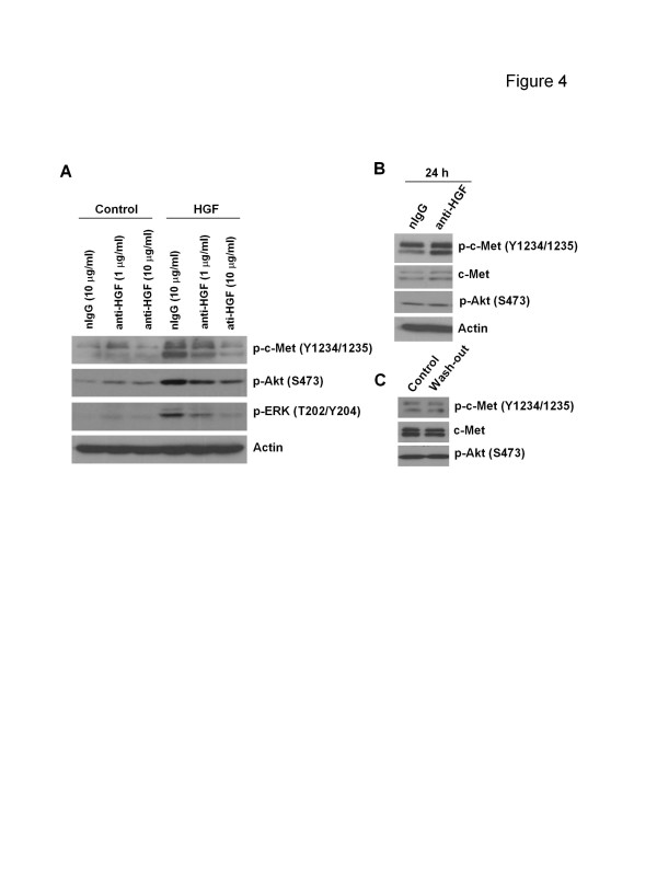
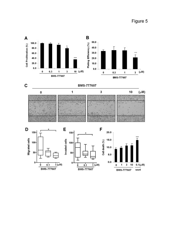
Although HGF mRNA could be detected in PC-3 cells, the molecular weight of secreted "HGF" protein was inconsistent with the functional recombinant HGF. Furthermore, conditioned medium from PC-3 cell cultures was ineffective at triggering either motogenic behavior or c-Met signaling in DU145, another prostate cancer cell line expressing c-Met but lacking basal c-Met activation. PC-3 cells also were not responsive to the anti-HGF neutralizing antibody in experiments assessing proliferation, migration, or c-Met signaling. BMS-777607 treatment with micromolar doses nonetheless led to significant inhibition of multiple PC-3 cell functions including proliferation, clonogenicity, migration and invasion. At the molecular level, BMS-777607 suppressed autophosphorylated c-Met and downstream c-Src and Akt pathways.
These results suggest that the constitutive c-Met activation in PC-3 is independent of autocrine stimulation. Because PC-3 cells were responsive to BMS-777607 but not the anti-HGF antibody, the findings also indicate that under circumstances where c-Met is constitutively hyperactive in the absence of functional HGF, targeting the c-Met receptor remains a viable therapeutic option to impede cancer progression.
c-Met 受体酪氨酸激酶在许多实体瘤中异常激活。在之前的研究中,我们表明前列腺癌细胞 PC-3 表现出组成型激活的 c-Met,而没有外源性肝细胞生长因子(HGF);然而,这种特征是否归因于内源性 HGF/c-Met 自分泌环仍存在争议。在当前的研究中,我们检查了 PC-3 细胞对抗 HGF 中和抗体或小分子 Met 激酶抑制剂(BMS-777607)的反应。
通过监测 HGF 刺激后细胞形态来测试细胞散射。通过“划痕愈合”和 Transwell 分析检测细胞迁移,通过 Matrigel 包被的 Transwell 分析检测侵袭。通过 MTT、集落形成和台盼蓝排除试验分别测定增殖、存活和失巢凋亡。通过实时 PCR 和 Western blot 分别评估基因和蛋白质表达。
尽管可以在 PC-3 细胞中检测到 HGF mRNA,但分泌的“HGF”蛋白的分子量与功能性重组 HGF 不一致。此外,来自 PC-3 细胞培养物的条件培养基在触发另一种前列腺癌细胞系 DU145 的运动行为或 c-Met 信号传导方面均无效,该细胞系表达 c-Met,但缺乏基础 c-Met 激活。在评估增殖、迁移或 c-Met 信号传导的实验中,PC-3 细胞也对抗 HGF 中和抗体无反应。然而,用微摩尔剂量的 BMS-777607 治疗会导致包括增殖、集落形成、迁移和侵袭在内的多种 PC-3 细胞功能的显著抑制。在分子水平上,BMS-777607 抑制了自磷酸化的 c-Met 和下游的 c-Src 和 Akt 途径。
这些结果表明,PC-3 中的组成型 c-Met 激活独立于自分泌刺激。由于 PC-3 细胞对 BMS-777607 有反应,但对抗 HGF 抗体无反应,这些发现还表明,在没有功能性 HGF 的情况下 c-Met 持续过度活跃的情况下,靶向 c-Met 受体仍然是阻止癌症进展的可行治疗选择。