• 文献检索
  • 文档翻译
  • 深度研究
  • 学术资讯
  • Suppr Zotero 插件Zotero 插件
  • 邀请有礼
  • 套餐&价格
  • 历史记录
应用&插件
Suppr Zotero 插件Zotero 插件浏览器插件Mac 客户端Windows 客户端微信小程序
定价
高级版会员购买积分包购买API积分包
服务
文献检索文档翻译深度研究API 文档MCP 服务
关于我们
关于 Suppr公司介绍联系我们用户协议隐私条款
关注我们

Suppr 超能文献

核心技术专利:CN118964589B侵权必究
粤ICP备2023148730 号-1Suppr @ 2026

文献检索

告别复杂PubMed语法,用中文像聊天一样搜索,搜遍4000万医学文献。AI智能推荐,让科研检索更轻松。

立即免费搜索

文件翻译

保留排版,准确专业,支持PDF/Word/PPT等文件格式,支持 12+语言互译。

免费翻译文档

深度研究

AI帮你快速写综述,25分钟生成高质量综述,智能提取关键信息,辅助科研写作。

立即免费体验

在细胞黏附过程中,顶端富含肌动蛋白的结构域驱动细胞极性的建立。

An apical actin-rich domain drives the establishment of cell polarity during cell adhesion.

机构信息

Dipartimento di Biotecnologie, Università degli Studi di Siena, Via Fiorentina 1, 53100 Siena, Italy.

出版信息

Histochem Cell Biol. 2012 Sep;138(3):419-33. doi: 10.1007/s00418-012-0965-9. Epub 2012 May 29.

DOI:10.1007/s00418-012-0965-9
PMID:22644377
原文链接:https://pmc.ncbi.nlm.nih.gov/articles/PMC3426669/
Abstract

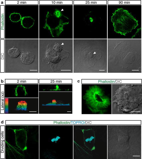
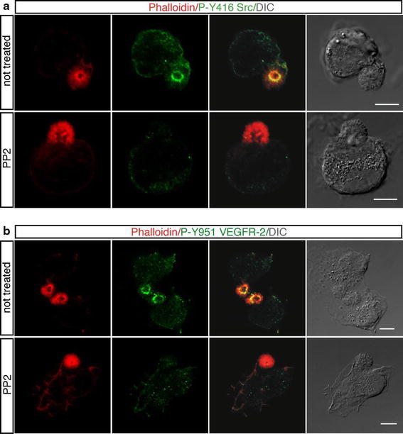
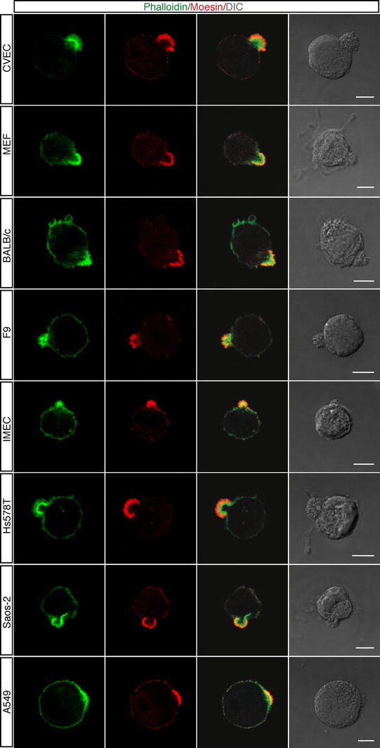
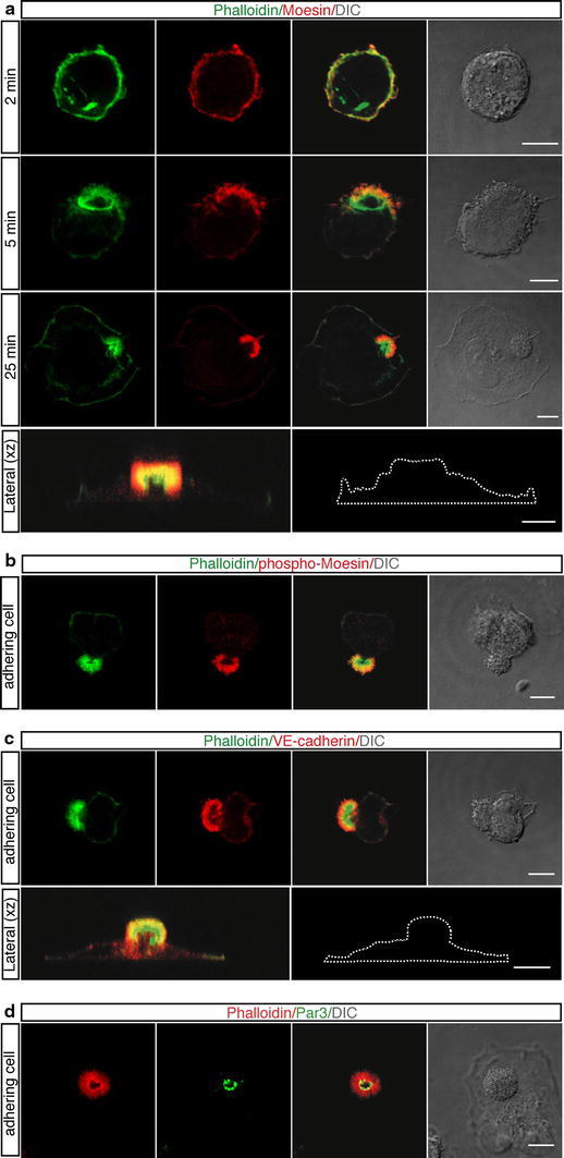
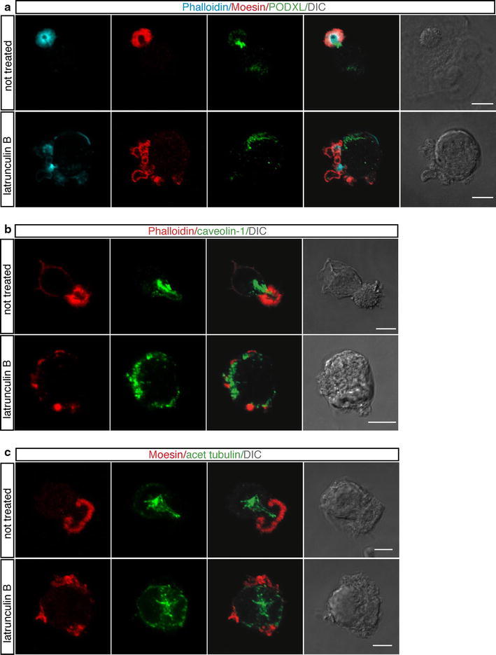
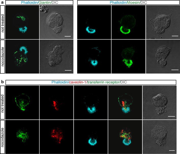
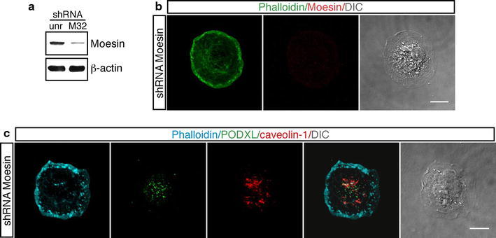
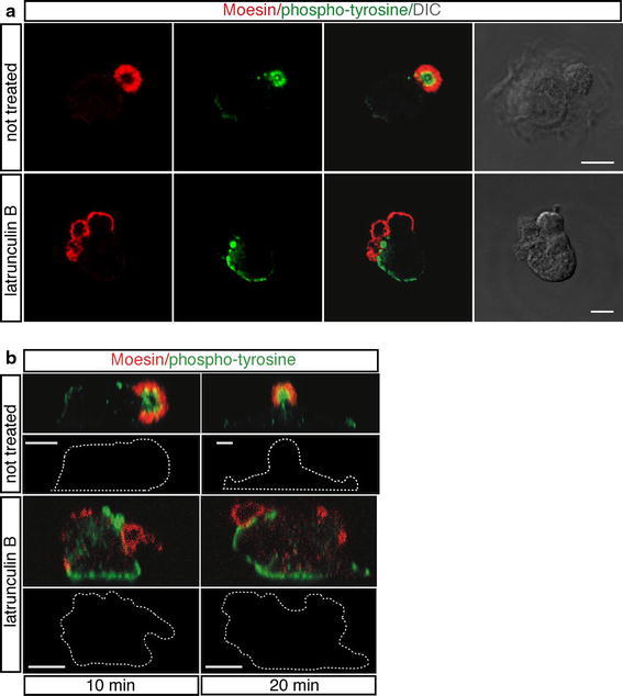
One of the most important questions in cell biology concerns how cells reorganize after sensing polarity cues. In the present study, we describe the formation of an actin-rich domain on the apical surface of human primary endothelial cells adhering to the substrate and investigate its role in cell polarity. We used confocal immunofluorescence procedures to follow the redistribution of proteins required for endothelial cell polarity during spreading initiation. Activated Moesin, vascular endothelial cadherin and partitioning defective 3 were found to be localized in the apical domain, whereas podocalyxin and caveolin-1 were distributed along the microtubule cytoskeleton axis, oriented from the centrosome to the cortical actin-rich domain. Moreover, activated signaling molecules were localized in the core of the apical domain in tight association with filamentous actin. During cell attachment, loss of the apical domain by Moesin silencing or drug disruption of the actin cytoskeleton caused irregular cell spreading and mislocalization of polarity markers. In conclusion, our results suggest that the apical domain that forms during the spreading process is a structural organizer of cell polarity by regulating trafficking and activation of signaling proteins.

摘要

细胞生物学中最重要的问题之一是细胞在感知极性线索后如何重新组织。在本研究中,我们描述了人原代内皮细胞在附着于底物时在顶端表面形成富含肌动蛋白的域,并研究了其在细胞极性中的作用。我们使用共聚焦免疫荧光程序来跟踪在起始扩展过程中内皮细胞极性所需的蛋白质的重分布。发现活化的 Moesin、血管内皮钙黏蛋白和分配缺陷 3 定位于顶端域,而足细胞蛋白和窖蛋白 1 沿微管细胞骨架轴分布,从中心体到富含肌动蛋白的皮质域定向。此外,活化的信号分子与丝状肌动蛋白紧密结合定位于顶端域的核心。在细胞附着过程中,通过 Moesin 沉默或药物破坏肌动蛋白细胞骨架损失顶端域导致不规则的细胞扩展和极性标记物的定位错误。总之,我们的结果表明,在扩展过程中形成的顶端域通过调节信号蛋白的运输和激活来充当细胞极性的结构组织者。

https://cdn.ncbi.nlm.nih.gov/pmc/blobs/2115/3426669/840b029115bb/418_2012_965_Fig8_HTML.jpg
https://cdn.ncbi.nlm.nih.gov/pmc/blobs/2115/3426669/27f84fafec90/418_2012_965_Fig1_HTML.jpg
https://cdn.ncbi.nlm.nih.gov/pmc/blobs/2115/3426669/8602849f080e/418_2012_965_Fig2_HTML.jpg
https://cdn.ncbi.nlm.nih.gov/pmc/blobs/2115/3426669/4d3689e919e7/418_2012_965_Fig3_HTML.jpg
https://cdn.ncbi.nlm.nih.gov/pmc/blobs/2115/3426669/7565f3265f38/418_2012_965_Fig4_HTML.jpg
https://cdn.ncbi.nlm.nih.gov/pmc/blobs/2115/3426669/42cb0ee01f79/418_2012_965_Fig5_HTML.jpg
https://cdn.ncbi.nlm.nih.gov/pmc/blobs/2115/3426669/ca44693bf584/418_2012_965_Fig6_HTML.jpg
https://cdn.ncbi.nlm.nih.gov/pmc/blobs/2115/3426669/309e26003f3c/418_2012_965_Fig7_HTML.jpg
https://cdn.ncbi.nlm.nih.gov/pmc/blobs/2115/3426669/840b029115bb/418_2012_965_Fig8_HTML.jpg

相似文献

1
An apical actin-rich domain drives the establishment of cell polarity during cell adhesion.在细胞黏附过程中,顶端富含肌动蛋白的结构域驱动细胞极性的建立。
Histochem Cell Biol. 2012 Sep;138(3):419-33. doi: 10.1007/s00418-012-0965-9. Epub 2012 May 29.
2
DAPLE orchestrates apical actomyosin assembly from junctional polarity complexes.DAPLE 从连接极性复合物中协调顶端肌动球蛋白的组装。
J Cell Biol. 2022 May 2;221(5). doi: 10.1083/jcb.202111002. Epub 2022 Apr 7.
3
Moesin functions antagonistically to the Rho pathway to maintain epithelial integrity.膜突蛋白对Rho信号通路起拮抗作用以维持上皮完整性。
Nature. 2003 Jan 2;421(6918):83-7. doi: 10.1038/nature01295.
4
Rho GTPase controls Drosophila salivary gland lumen size through regulation of the actin cytoskeleton and Moesin.Rho GTPase 通过调控肌动蛋白细胞骨架和 Moesin 控制果蝇唾液腺管腔大小。
Development. 2011 Dec;138(24):5415-27. doi: 10.1242/dev.069831. Epub 2011 Nov 9.
5
Role for actin filament turnover and a myosin II motor in cytoskeleton-driven disassembly of the epithelial apical junctional complex.肌动蛋白丝周转和肌球蛋白II马达在上皮顶端连接复合体的细胞骨架驱动解体中的作用。
Mol Biol Cell. 2004 Jun;15(6):2639-51. doi: 10.1091/mbc.e04-02-0163. Epub 2004 Mar 26.
6
A CD317/tetherin-RICH2 complex plays a critical role in the organization of the subapical actin cytoskeleton in polarized epithelial cells.CD317/栓系蛋白-RICH2复合物在极化上皮细胞的顶端下肌动蛋白细胞骨架组织中起关键作用。
J Cell Biol. 2009 Mar 9;184(5):721-36. doi: 10.1083/jcb.200804154.
7
Spatial control of actin organization at adherens junctions by a synaptotagmin-like protein Btsz.一种类突触结合蛋白Btsz对黏着连接中肌动蛋白组织的空间控制。
Nature. 2006 Aug 3;442(7102):580-4. doi: 10.1038/nature04935. Epub 2006 Jul 9.
8
Moesin orchestrates cortical polarity of melanoma tumour cells to initiate 3D invasion.埃兹蛋白调控黑色素瘤肿瘤细胞的皮质极性以启动三维侵袭。
J Cell Sci. 2009 Oct 1;122(Pt 19):3492-501. doi: 10.1242/jcs.053157. Epub 2009 Sep 1.
9
High levels of E-/P-cadherin: correlation with decreased apical polarity of Na/K ATPase in bovine RPE cells in situ.E-钙黏蛋白/上皮钙黏蛋白的高表达:与牛视网膜色素上皮细胞原位钠钾ATP酶顶端极性降低的相关性
Invest Ophthalmol Vis Sci. 2000 Jun;41(7):1945-52.
10
Constitutive activation of Rho proteins by CNF-1 influences tight junction structure and epithelial barrier function.CNF-1对Rho蛋白的组成性激活会影响紧密连接结构和上皮屏障功能。
J Cell Sci. 2003 Feb 15;116(Pt 4):725-42. doi: 10.1242/jcs.00300.

引用本文的文献

1
CD93 in Health and Disease: Bridging Physiological Functions and Clinical Applications.健康与疾病中的CD93:连接生理功能与临床应用
Int J Mol Sci. 2025 Sep 4;26(17):8617. doi: 10.3390/ijms26178617.
2
Semi-synthetic fibrous fibrin composites promote 3D microvascular assembly, survival, and host integration of endothelial cells without mesenchymal cell support.半合成纤维蛋白复合材料可促进内皮细胞的三维微血管组装、存活及宿主整合,且无需间充质细胞支持。
Bioact Mater. 2025 Apr 3;49:652-669. doi: 10.1016/j.bioactmat.2025.02.029. eCollection 2025 Jul.
3
Shroom3 facilitates optic fissure closure via tissue alignment and reestablishment of apical-basal polarity during epithelial fusion.

本文引用的文献

1
Molecular control of endothelial cell behaviour during blood vessel morphogenesis.血管生成过程中内皮细胞行为的分子调控。
Nat Rev Mol Cell Biol. 2011 Aug 23;12(9):551-64. doi: 10.1038/nrm3176.
2
Attachment of HeLa cells during early G1 phase.早 G1 期 HeLa 细胞的附着。
Histochem Cell Biol. 2011 Oct;136(4):399-411. doi: 10.1007/s00418-011-0852-9. Epub 2011 Aug 11.
3
Ezrin, Radixin and Moesin: key regulators of membrane-cortex interactions and signaling.埃兹蛋白、根蛋白和膜突蛋白:调节质膜-细胞皮质连接及信号转导的关键分子。
Shroom3通过上皮融合过程中的组织排列和顶-基极性的重建促进视裂闭合。
Dev Biol. 2025 Jun;522:91-105. doi: 10.1016/j.ydbio.2025.03.008. Epub 2025 Mar 18.
4
Role of CD93 in Health and Disease.CD93 在健康和疾病中的作用。
Cells. 2023 Jul 4;12(13):1778. doi: 10.3390/cells12131778.
5
Myo-Inositol Reverses TGF-β1-Induced EMT in MCF-10A Non-Tumorigenic Breast Cells.肌醇可逆转转化生长因子-β1诱导的MCF-10A非致瘤性乳腺细胞中的上皮-间质转化。
Cancers (Basel). 2023 Apr 15;15(8):2317. doi: 10.3390/cancers15082317.
6
Actin cytoskeleton in angiogenesis.血管生成中的肌动蛋白细胞骨架。
Biol Open. 2022 Dec 15;11(12). doi: 10.1242/bio.058899. Epub 2022 Nov 29.
7
The Relative Densities of Cytoplasm and Nuclear Compartments Are Robust against Strong Perturbation.细胞质和核区室的相对密度对强烈扰动具有稳健性。
Biophys J. 2020 Nov 17;119(10):1946-1957. doi: 10.1016/j.bpj.2020.08.044. Epub 2020 Oct 20.
8
The Binding of CD93 to Multimerin-2 Promotes Choroidal Neovascularization.CD93 与多配体蛋白聚糖-2 的结合促进脉络膜新生血管形成。
Invest Ophthalmol Vis Sci. 2020 Jul 1;61(8):30. doi: 10.1167/iovs.61.8.30.
9
Hepatocyte growth factor-regulated tyrosine kinase substrate is essential for endothelial cell polarity and cerebrovascular stability.肝细胞生长因子调节的酪氨酸激酶底物对于内皮细胞极性和脑血管稳定性至关重要。
Cardiovasc Res. 2021 Jan 21;117(2):533-546. doi: 10.1093/cvr/cvaa016.
10
Histochemistry and Cell Biology: 61 years and not tired at all.《组织化学与细胞生物学》:61 年,活力依旧。
Histochem Cell Biol. 2019 Jul;152(1):1-11. doi: 10.1007/s00418-019-01796-2.
Curr Opin Cell Biol. 2011 Aug;23(4):377-82. doi: 10.1016/j.ceb.2011.04.011. Epub 2011 May 16.
4
The posttranslational modification of tubulin undergoes a switch from detyrosination to acetylation as epithelial cells become polarized.微管蛋白的翻译后修饰在上皮细胞极化时经历从脱酪氨酸化到乙酰化的转变。
Mol Biol Cell. 2011 Apr;22(7):1045-57. doi: 10.1091/mbc.E10-06-0519. Epub 2011 Feb 9.
5
Moesin1 and Ve-cadherin are required in endothelial cells during in vivo tubulogenesis.Moesin1 和 Ve-cadherin 在体内小管形成过程中内皮细胞中是必需的。
Development. 2010 Sep;137(18):3119-28. doi: 10.1242/dev.048785.
6
Cell polarity in eggs and epithelia: parallels and diversity.卵子和上皮细胞中的细胞极性:相似性与多样性。
Cell. 2010 May 28;141(5):757-74. doi: 10.1016/j.cell.2010.05.011.
7
A complex between FAK, RACK1, and PDE4D5 controls spreading initiation and cancer cell polarity.FAK、RACK1 和 PDE4D5 复合物控制着细胞铺展的起始和癌细胞极性。
Curr Biol. 2010 Jun 22;20(12):1086-92. doi: 10.1016/j.cub.2010.04.042. Epub 2010 May 20.
8
Trypsin-induced proteome alteration during cell subculture in mammalian cells.胰蛋白酶诱导的哺乳动物细胞传代培养过程中的蛋白质组改变。
J Biomed Sci. 2010 May 11;17(1):36. doi: 10.1186/1423-0127-17-36.
9
Endothelial cell adhesion to the extracellular matrix induces c-Src-dependent VEGFR-3 phosphorylation without the activation of the receptor intrinsic kinase activity.内皮细胞黏附于细胞外基质会诱导 c-Src 依赖性 VEGFR-3 磷酸化,而不会激活受体内在激酶活性。
Circ Res. 2010 Jun 25;106(12):1839-48. doi: 10.1161/CIRCRESAHA.109.206326. Epub 2010 Apr 29.
10
CCM1 regulates vascular-lumen organization by inducing endothelial polarity.CCM1 通过诱导内皮细胞极性来调节血管腔的组织。
J Cell Sci. 2010 Apr 1;123(Pt 7):1073-80. doi: 10.1242/jcs.059329.