Department of Developmental Dentistry, The University of Texas Health Science Center at San Antonio, San Antonio, TX 78229-3900, USA.
J Mol Histol. 2012 Oct;43(5):473-85. doi: 10.1007/s10735-012-9423-1. Epub 2012 May 31.
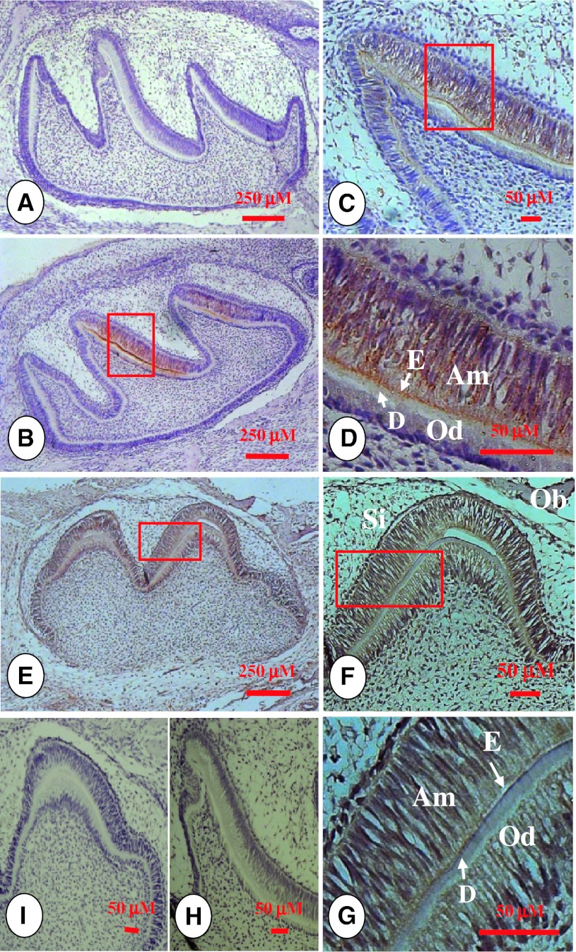
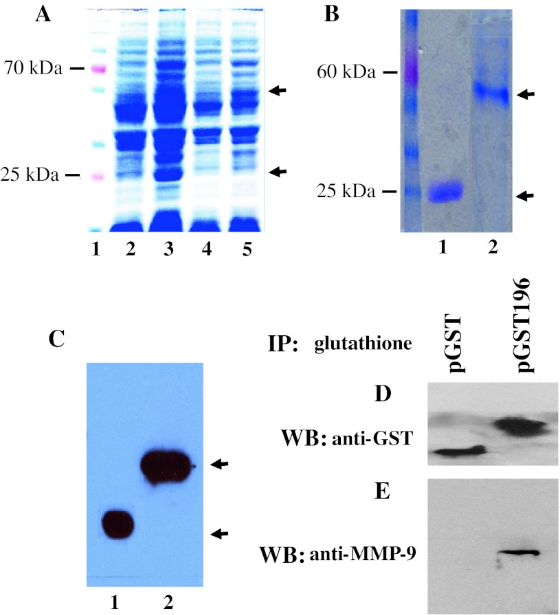
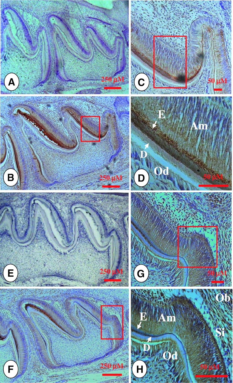
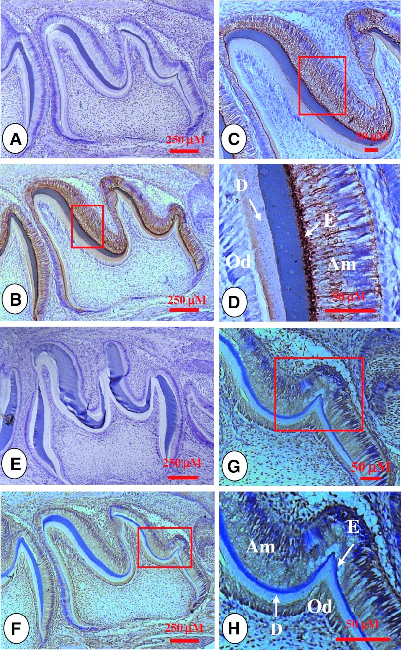
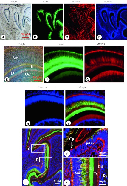
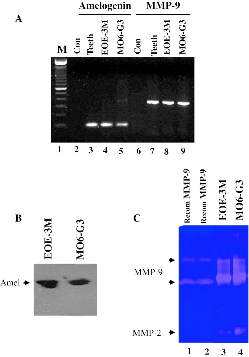
Amelogenin is the most abundant matrix protein in enamel. Proper amelogenin processing by proteinases is necessary for its biological functions during amelogenesis. Matrix metalloproteinase 9 (MMP-9) is responsible for the turnover of matrix components. The relationship between MMP-9 and amelogenin during tooth development remains unknown. We tested the hypothesis that MMP-9 binds to amelogenin and they are co-expressed in ameloblasts during amelogenesis. We evaluated the distribution of both proteins in the mouse teeth using immunohistochemistry and confocal microscopy. At postnatal day 2, the spatial distribution of amelogenin and MMP-9 was co-localized in preameloblasts, secretory ameloblasts, enamel matrix and odontoblasts. At the late stages of mouse tooth development, expression patterns of amelogenin and MMP-9 were similar to that seen in postnatal day 2. Their co-expression was further confirmed by RT-PCR, Western blot and enzymatic zymography analyses in enamel organ epithelial and odontoblast-like cells. Immunoprecipitation assay revealed that MMP-9 binds to amelogenin. The MMP-9 cleavage sites in amelogenin proteins across species were found using bio-informative software program. Analyses of these data suggest that MMP-9 may be involved in controlling amelogenin processing and enamel formation.
釉原蛋白是釉质中最丰富的基质蛋白。蛋白酶对釉原蛋白的适当处理对于其在釉质发生过程中的生物学功能是必要的。基质金属蛋白酶 9(MMP-9)负责基质成分的更新。MMP-9 与牙发育过程中釉原蛋白之间的关系尚不清楚。我们检验了以下假设:MMP-9 与釉原蛋白结合,并且它们在釉质发生过程中在成釉细胞中共同表达。我们使用免疫组织化学和共聚焦显微镜评估了这两种蛋白质在小鼠牙齿中的分布。在出生后第 2 天,釉原蛋白和 MMP-9 的空间分布在成釉前体细胞、分泌型成釉细胞、釉基质和成牙本质细胞中共同定位。在小鼠牙齿发育的后期阶段,釉原蛋白和 MMP-9 的表达模式与出生后第 2 天相似。在釉器官上皮细胞和成牙本质细胞样细胞中通过 RT-PCR、Western blot 和酶谱分析进一步证实了它们的共表达。免疫沉淀分析显示 MMP-9 与釉原蛋白结合。使用生物信息学软件程序在跨物种的釉原蛋白中发现了 MMP-9 的切割位点。对这些数据的分析表明,MMP-9 可能参与控制釉原蛋白的处理和牙釉质的形成。