Orthopedic Institute of Chinese People's Liberation Army, Xijing Hospital, Fourth Military Medical University, Xi'an, Shanxi, China.
Arch Med Sci. 2012 May 9;8(2):199-208. doi: 10.5114/aoms.2012.28545.
With the increase in joint revision surgery after arthroplasty, defects of hydroxyapatite (HA)-coated prostheses have been observed increasingly often. These defects adversely affect the prosthetic stability in vivo. This study has analyzed the potential effect of the adhesive strength of HA coating on the stability of HA-coated prostheses in vivo after its implantation.
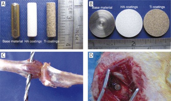
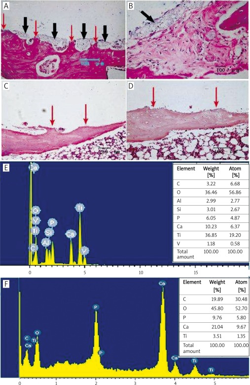
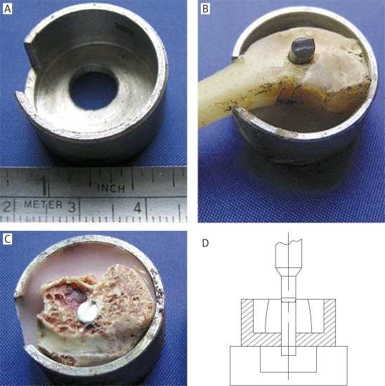
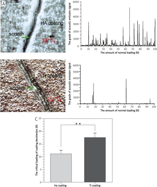
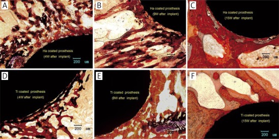
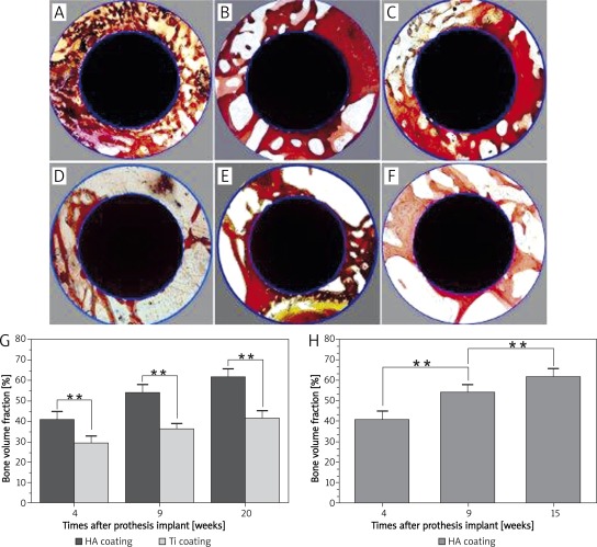
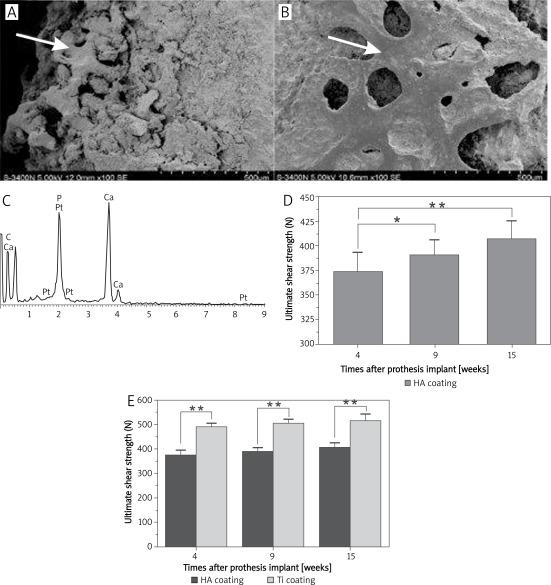
Sixty experimental rabbits were divided into HA- and Ti-coated groups. HA-coated prostheses were implanted into the bilateral epicondyle of rabbits femurs. Ti-coated prostheses were implanted as control. At different time points(4, 9, and 15 weeks) after implantation, bone tissue samples were fetched out respectively for histomorphometric analysis. Push-out testing was used to detect the ultimate shear strength at the bone-prosthesis interface. Scanning electron microscope (SEM) observation and energy-dispersive X-ray spectroscopy (EDX) analysis were used to observe the changes in surface composition of the prostheses after the ultimate shear strength testing. The coating adhesive strength of two kinds of coatings were also examined by scratch testing.
Hydroxyapatite coating has an obvious advantage in facilitating osteogenesis and its plays a critical role in the stability of prostheses. However, the ultimate shear strength of HA-coated prostheses is much lower than that of Ti-coated implants (p < 0.01). Further study has demonstrated that the stability of HA-coated prostheses in vivo is affected by the relatively low adhesive strength between coating and substrate.
Obvious advantage in facilitating osteogenesis around HA-coated prostheses is not the only factor that determines the stability of prostheses in vivo.
随着关节翻修手术后的增加,羟基磷灰石(HA)涂层假体的缺陷越来越多地被观察到。这些缺陷会对假体在体内的稳定性产生不利影响。本研究分析了 HA 涂层的粘附强度对植入后体内 HA 涂层假体稳定性的潜在影响。
60 只实验兔分为 HA 和 Ti 涂层组。HA 涂层假体植入兔股骨髁双侧。Ti 涂层假体作为对照植入。植入后 4、9 和 15 周时,分别取出骨组织样本进行组织形态计量学分析。采用推出试验检测骨-假体界面的极限抗剪强度。扫描电子显微镜(SEM)观察和能谱分析(EDX)用于观察极限抗剪强度试验后假体表面成分的变化。还通过划痕试验检查了两种涂层的涂层粘附强度。
HA 涂层在促进成骨方面具有明显优势,对假体的稳定性起着关键作用。然而,HA 涂层假体的极限抗剪强度明显低于 Ti 涂层植入物(p < 0.01)。进一步的研究表明,体内 HA 涂层假体的稳定性受到涂层与基底之间相对较低的粘附强度的影响。
HA 涂层假体周围明显的促进成骨优势并不是决定假体在体内稳定性的唯一因素。