• 文献检索
  • 文档翻译
  • 深度研究
  • 学术资讯
  • Suppr Zotero 插件Zotero 插件
  • 邀请有礼
  • 套餐&价格
  • 历史记录
应用&插件
Suppr Zotero 插件Zotero 插件浏览器插件Mac 客户端Windows 客户端微信小程序
定价
高级版会员购买积分包购买API积分包
服务
文献检索文档翻译深度研究API 文档MCP 服务
关于我们
关于 Suppr公司介绍联系我们用户协议隐私条款
关注我们

Suppr 超能文献

核心技术专利:CN118964589B侵权必究
粤ICP备2023148730 号-1Suppr @ 2026

文献检索

告别复杂PubMed语法,用中文像聊天一样搜索,搜遍4000万医学文献。AI智能推荐,让科研检索更轻松。

立即免费搜索

文件翻译

保留排版,准确专业,支持PDF/Word/PPT等文件格式,支持 12+语言互译。

免费翻译文档

深度研究

AI帮你快速写综述,25分钟生成高质量综述,智能提取关键信息,辅助科研写作。

立即免费体验

骨盆骨肿瘤的影像学综述——第一部分:骨盆良性肿瘤

Imaging review of skeletal tumors of the pelvis--part I: benign tumors of the pelvis.

作者信息

Girish Gandikota, Finlay Karen, Morag Yoav, Brandon Catherine, Jacobson Jon, Jamadar David

机构信息

Department of Radiology, University of Michigan, 1500 E. Medical Center Drive, TC-2910, Ann Arbor, MI 48109-0326, USA.

出版信息

ScientificWorldJournal. 2012;2012:290930. doi: 10.1100/2012/290930. Epub 2012 May 15.

DOI:10.1100/2012/290930
PMID:22666102
原文链接:https://pmc.ncbi.nlm.nih.gov/articles/PMC3362015/
Abstract

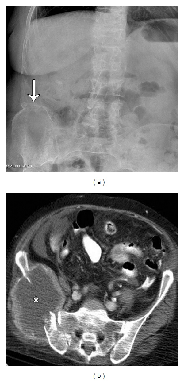
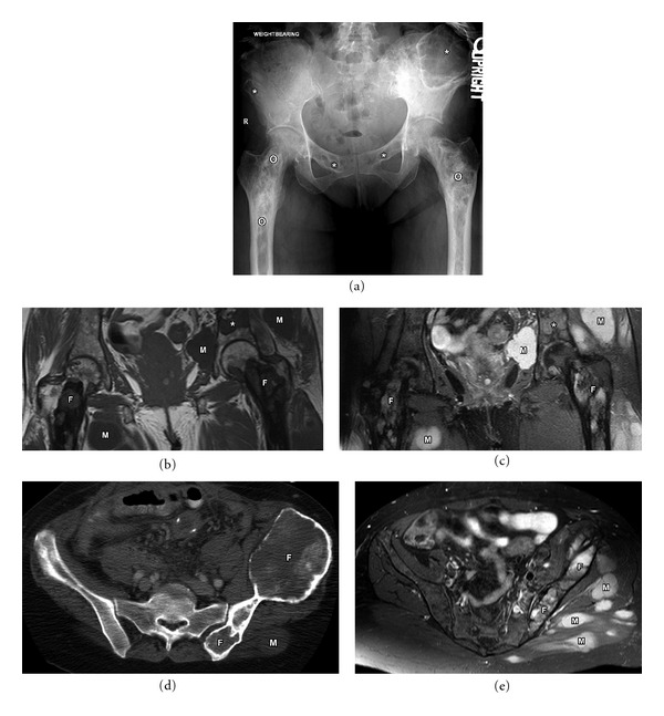
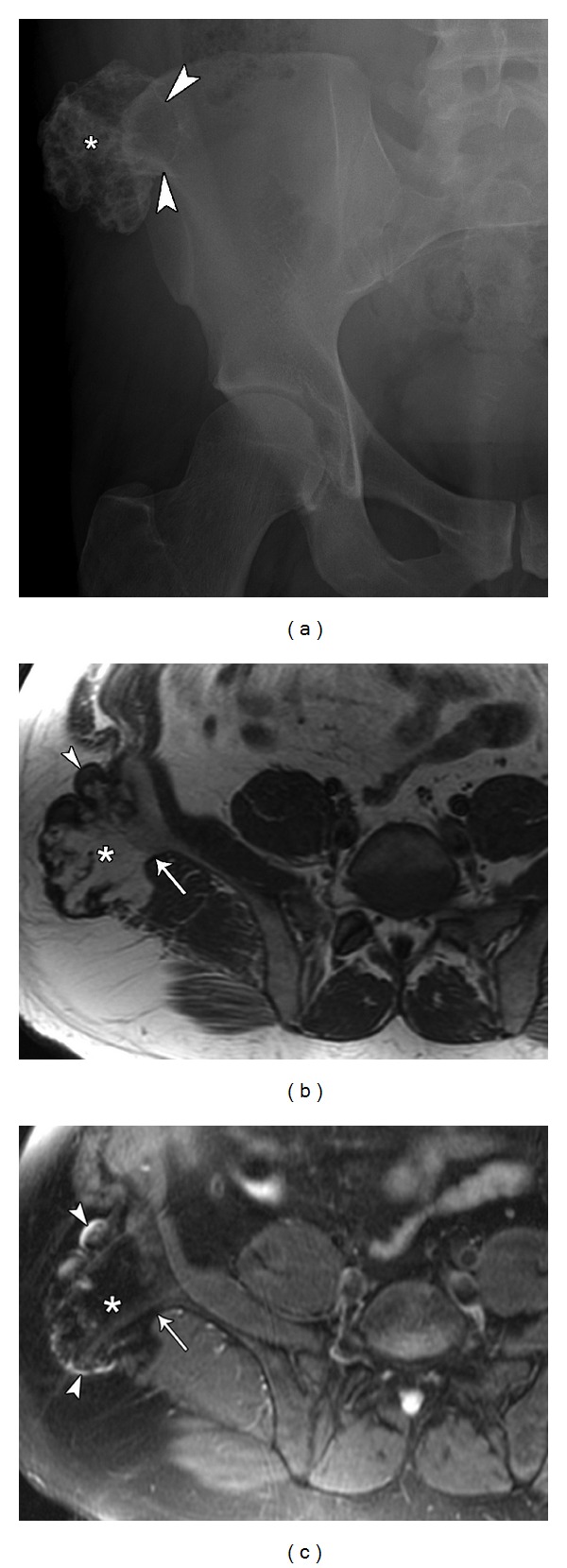
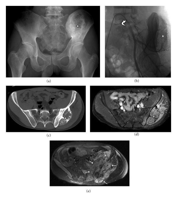
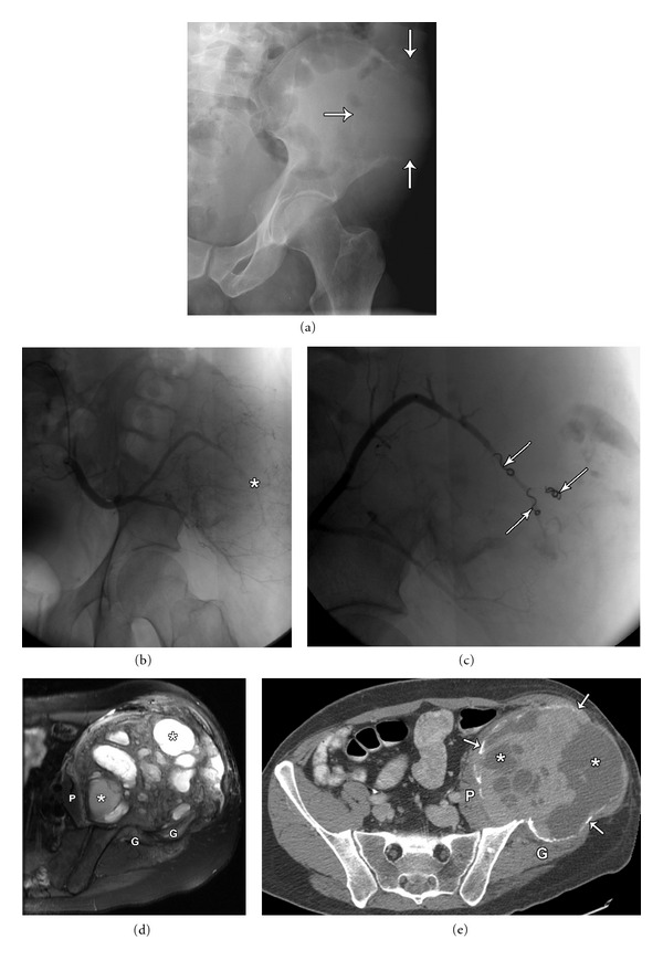
The osseous pelvis is a well-recognized site of origin of numerous primary and secondary musculoskeletal tumors. The radiologic evaluation of a pelvic lesion often begins with the plain film and proceeds to computed tomography (CT), or magnetic resonance imaging (MRI) and possibly biopsy. Each of these modalities, with inherent advantages and disadvantages, has a role in the workup of pelvic osseous masses. Clinical history and imaging characteristics can significantly narrow the broad differential diagnosis for osseous pelvic lesions. The purpose of this review is to familiarize the radiologist with the presentation and appearance of some of the common benign neoplasms of the osseous pelvis and share our experience and approach in diagnosing these lesions.

摘要

骨盆是众多原发性和继发性肌肉骨骼肿瘤公认的起源部位。对骨盆病变的放射学评估通常从平片开始,然后进行计算机断层扫描(CT)、磁共振成像(MRI),可能还包括活检。这些检查方法各有其固有的优缺点,在骨盆骨质肿块的检查中都发挥着作用。临床病史和影像学特征能够显著缩小骨盆骨质病变广泛的鉴别诊断范围。本综述的目的是让放射科医生熟悉骨盆常见良性肿瘤的表现及影像学特征,并分享我们在诊断这些病变方面的经验和方法。

https://cdn.ncbi.nlm.nih.gov/pmc/blobs/9388/3362015/34c46ccc933c/TSWJ2012-290930.005.jpg
https://cdn.ncbi.nlm.nih.gov/pmc/blobs/9388/3362015/35294bca9cdb/TSWJ2012-290930.001.jpg
https://cdn.ncbi.nlm.nih.gov/pmc/blobs/9388/3362015/2f7162a4c374/TSWJ2012-290930.002.jpg
https://cdn.ncbi.nlm.nih.gov/pmc/blobs/9388/3362015/86babe788ebc/TSWJ2012-290930.003.jpg
https://cdn.ncbi.nlm.nih.gov/pmc/blobs/9388/3362015/a5e20db195c0/TSWJ2012-290930.004.jpg
https://cdn.ncbi.nlm.nih.gov/pmc/blobs/9388/3362015/34c46ccc933c/TSWJ2012-290930.005.jpg

相似文献

1
Imaging review of skeletal tumors of the pelvis--part I: benign tumors of the pelvis.骨盆骨肿瘤的影像学综述——第一部分:骨盆良性肿瘤
ScientificWorldJournal. 2012;2012:290930. doi: 10.1100/2012/290930. Epub 2012 May 15.
2
Imaging review of skeletal tumors of the pelvis malignant tumors and tumor mimics.骨盆骨肿瘤的影像学回顾:恶性肿瘤及肿瘤样病变
ScientificWorldJournal. 2012;2012:240281. doi: 10.1100/2012/240281. Epub 2012 Apr 19.
3
Hip and Pelvis Bone Tumors: Can You Make It Simple?
Semin Musculoskelet Radiol. 2019 Jun;23(3):e37-e57. doi: 10.1055/s-0039-1684023. Epub 2019 Jun 4.
4
The role of diffusion-weighted MRI (DWI) in the differentiation of benign from malignant skeletal lesions of the pelvis.扩散加权磁共振成像(DWI)在骨盆骨骼良性与恶性病变鉴别中的作用
Eur J Radiol. 2016 Dec;85(12):2262-2268. doi: 10.1016/j.ejrad.2016.10.014. Epub 2016 Oct 17.
5
Benign osseous and articular abnormalities of the pelvis: a review of CT imaging findings.
Clin Imaging. 2015 Mar-Apr;39(2):186-93. doi: 10.1016/j.clinimag.2014.08.005. Epub 2014 Oct 16.
6
Magnetic Resonance Imaging of Pelvic Masses: A Compartmental Approach.盆腔肿块的磁共振成像:一种分区法
Semin Ultrasound CT MR. 2017 Jun;38(3):213-230. doi: 10.1053/j.sult.2016.11.004. Epub 2016 Nov 28.
7
Accuracy of chemical shift MR imaging in diagnosing indeterminate bone marrow lesions in the pelvis: review of a single institution's experience.化学位移磁共振成像在诊断骨盆中不确定骨髓病变的准确性:单机构经验回顾
Skeletal Radiol. 2014 Aug;43(8):1079-84. doi: 10.1007/s00256-014-1886-6. Epub 2014 Apr 30.
8
MRI of Tumors and Tumor Mimics in the Female Pelvis: Anatomic Pelvic Space-based Approach.女性盆腔肿瘤及肿瘤样病变的 MRI:基于解剖盆腔间隙的方法
Radiographics. 2019 Jul-Aug;39(4):1205-1229. doi: 10.1148/rg.2019180173.
9
The current role of radiography in the assessment of skeletal tumors and tumor-like lesions.放射成像在骨骼肿瘤及肿瘤样病变评估中的当前作用。
Eur J Radiol. 1998 May;27 Suppl 1:S77-85. doi: 10.1016/s0720-048x(98)00047-3.
10
Imaging of pseudoneoplastic masses associated with allografts.
Skeletal Radiol. 2007 Aug;36(8):747-53. doi: 10.1007/s00256-007-0292-8. Epub 2007 Apr 6.

引用本文的文献

1
Three-Dimensional Multimodality Image Reconstruction as Teaching Tool for Case-based learning among medical postgraduates: a focus on primary pelvic bone Tumour Education.三维多模态图像重建作为医学研究生基于病例学习的教学工具:以原发性骨盆骨肿瘤教学为重点。
BMC Med Educ. 2023 Dec 12;23(1):944. doi: 10.1186/s12909-023-04916-8.
2
Clinical Scenario and Imaging with Illustrations of Giant Cell Tumor of Bone: A Retrospective Analysis.骨巨细胞瘤的临床病例及影像学表现并附图:一项回顾性分析
Arch Bone Jt Surg. 2022 Jan;10(1):60-66. doi: 10.22038/ABJS.2021.50922.2522.
3
Axial Spondyloarthritis: Mimics and Pitfalls of Imaging Assessment.

本文引用的文献

1
PET/CT in primary musculoskeletal tumours: a step forward.正电子发射断层扫描/计算机断层扫描在原发性肌肉骨骼肿瘤中的应用:向前迈进了一步。
Eur Radiol. 2010 Dec;20(12):2959-72. doi: 10.1007/s00330-010-1862-z. Epub 2010 Jun 25.
2
The evolution of musculoskeletal tumor imaging.肌肉骨骼肿瘤影像学的发展
Radiol Clin North Am. 2009 May;47(3):435-53. doi: 10.1016/j.rcl.2008.12.002.
3
Dual-time point PET/CT with F-18 FDG for the differentiation of malignant and benign bone lesions.采用F-18氟代脱氧葡萄糖进行双时相PET/CT检查以鉴别骨病变的良恶性
轴性脊柱关节炎:影像学评估的模仿者与陷阱
Front Med (Lausanne). 2021 Apr 22;8:658538. doi: 10.3389/fmed.2021.658538. eCollection 2021.
4
Tumours of the talus - A pictorial review.距骨肿瘤——影像学综述
J Clin Orthop Trauma. 2020 May-Jun;11(3):410-416. doi: 10.1016/j.jcot.2020.03.021. Epub 2020 Apr 11.
5
Treatment of the aneurysmal bone cyst by percutaneous intracystic sclerotherapy using ethanol ninety five percent in children.采用95%乙醇经皮囊肿内硬化疗法治疗儿童动脉瘤样骨囊肿。
Int Orthop. 2018 Jun;42(6):1413-1419. doi: 10.1007/s00264-018-3841-y. Epub 2018 Feb 28.
6
Systematic approach to musculoskeletal benign tumors.肌肉骨骼系统良性肿瘤的系统诊疗方法
Int J Surg Oncol (N Y). 2017 Dec;2(11):e46. doi: 10.1097/IJ9.0000000000000046. Epub 2017 Nov 14.
7
[Conventional radiological diagnosis of benign none neoplasms].[良性非肿瘤性疾病的传统放射学诊断]
Orthopade. 2017 Jun;46(6):477-483. doi: 10.1007/s00132-017-3424-4.
Skeletal Radiol. 2009 May;38(5):451-8. doi: 10.1007/s00256-008-0643-0. Epub 2009 Feb 10.
4
Can diffusion-weighted imaging be used to differentiate benign from pathologic fractures? A meta-analysis.
Skeletal Radiol. 2008 Sep;37(9):791-5. doi: 10.1007/s00256-008-0503-y. Epub 2008 Jun 13.
5
In vivo proton spectroscopy of giant cell tumor of the bone.骨巨细胞瘤的体内质子波谱分析
AJR Am J Roentgenol. 2008 Feb;190(2):W133-9. doi: 10.2214/AJR.07.2802.
6
Characterization of musculoskeletal lesions on 3-T proton MR spectroscopy.3-T质子磁共振波谱对肌肉骨骼病变的特征分析
AJR Am J Roentgenol. 2007 Jun;188(6):1513-20. doi: 10.2214/AJR.06.0935.
7
PET/CT: form and function.正电子发射断层显像/计算机断层扫描(PET/CT):形态与功能
Radiology. 2007 Feb;242(2):360-85. doi: 10.1148/radiol.2422051113.
8
Cancer's molecular sweet tooth and the Warburg effect.癌症的分子“嗜甜”特性与瓦伯格效应。
Cancer Res. 2006 Sep 15;66(18):8927-30. doi: 10.1158/0008-5472.CAN-06-1501.
9
Aneurysmal bone cyst: a review of 150 patients.动脉瘤样骨囊肿:150例患者的回顾
J Clin Oncol. 2005 Sep 20;23(27):6756-62. doi: 10.1200/JCO.2005.15.255.
10
Imaging findings of fibrous dysplasia with histopathologic and intraoperative correlation.纤维发育不良的影像学表现与组织病理学及术中情况的相关性
AJR Am J Roentgenol. 2004 Jun;182(6):1389-98. doi: 10.2214/ajr.182.6.1821389.