• 文献检索
  • 文档翻译
  • 深度研究
  • 学术资讯
  • Suppr Zotero 插件Zotero 插件
  • 邀请有礼
  • 套餐&价格
  • 历史记录
应用&插件
Suppr Zotero 插件Zotero 插件浏览器插件Mac 客户端Windows 客户端微信小程序
定价
高级版会员购买积分包购买API积分包
服务
文献检索文档翻译深度研究API 文档MCP 服务
关于我们
关于 Suppr公司介绍联系我们用户协议隐私条款
关注我们

Suppr 超能文献

核心技术专利:CN118964589B侵权必究
粤ICP备2023148730 号-1Suppr @ 2026

文献检索

告别复杂PubMed语法,用中文像聊天一样搜索,搜遍4000万医学文献。AI智能推荐,让科研检索更轻松。

立即免费搜索

文件翻译

保留排版,准确专业,支持PDF/Word/PPT等文件格式,支持 12+语言互译。

免费翻译文档

深度研究

AI帮你快速写综述,25分钟生成高质量综述,智能提取关键信息,辅助科研写作。

立即免费体验

间质细胞与卵巢癌细胞的相互作用触发了促转移特性。

Mesenchymal cell interaction with ovarian cancer cells triggers pro-metastatic properties.

机构信息

Stem Cell and Microenvironment Laboratory, Weill Cornell Medical College in Qatar, Education City, Qatar Foundation, Doha, Qatar.

出版信息

PLoS One. 2012;7(5):e38340. doi: 10.1371/journal.pone.0038340. Epub 2012 May 30.

DOI:10.1371/journal.pone.0038340
PMID:22666502
原文链接:https://pmc.ncbi.nlm.nih.gov/articles/PMC3364218/
Abstract

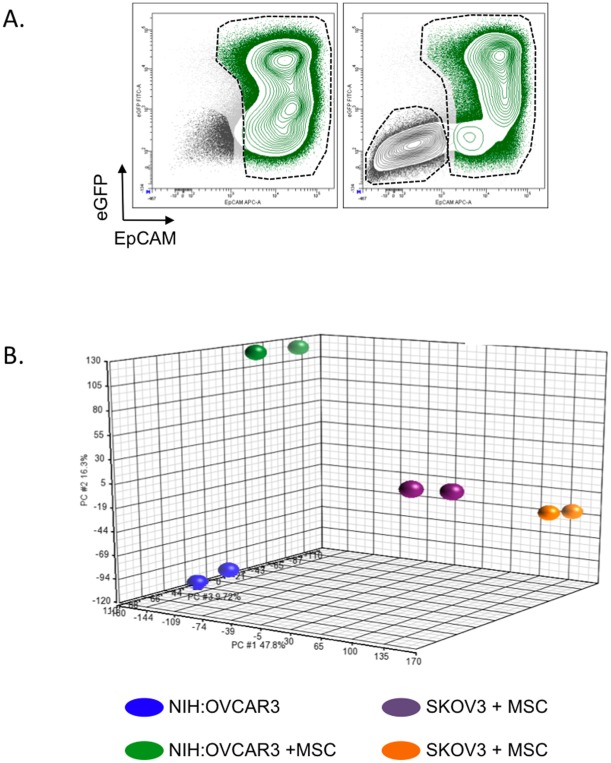
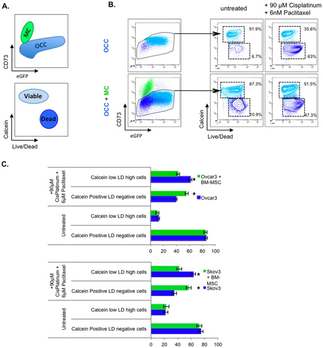
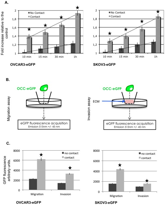
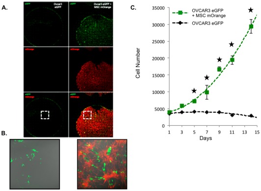
Tumor microenvironment is an important actor of ovarian cancer progression but the relations between mesenchymal cells and ovarian cancer cells remain unclear. The objective of this study was to determine the ovarian cancer cells' biological modifications induced by mesenchymal cells. To address this issue, we used two different ovarian cancer cell lines (NIH:OVCAR3 and SKOV3) and co-cultured them with mesenchymal cells. Upon co-culture the different cell populations were sorted to study their transcriptome and biological properties. Transcriptomic analysis revealed three biological-function gene clusters were enriched upon contact with mesenchymal cells. These were related to the increase of metastatic abilities (adhesion, migration and invasion), proliferation and chemoresistance in vitro. Therefore, contact with the mesenchymal cell niche could increase metastatic initiation and expansion through modification of cancer cells. Taken together these findings suggest that pathways involved in hetero-cellular interaction may be targeted to disrupt the acquired pro-metastatic profile.

摘要

肿瘤微环境是卵巢癌进展的重要因素,但间充质细胞与卵巢癌细胞之间的关系仍不清楚。本研究的目的是确定间充质细胞诱导的卵巢癌细胞的生物学改变。为了解决这个问题,我们使用了两种不同的卵巢癌细胞系(NIH:OVCAR3 和 SKOV3),并将它们与间充质细胞共培养。在共培养后,对不同的细胞群进行分选,以研究它们的转录组和生物学特性。转录组分析显示,与间充质细胞接触后富集了三个生物学功能基因簇。这些与体外转移能力(粘附、迁移和侵袭)、增殖和化疗耐药性的增加有关。因此,与间充质细胞龛的接触可能通过改变癌细胞来增加转移起始和扩张。总之,这些发现表明,涉及异细胞相互作用的途径可能成为靶向治疗以破坏获得的促转移表型的靶点。

https://cdn.ncbi.nlm.nih.gov/pmc/blobs/1ec7/3364218/88cdfc4b7463/pone.0038340.g004.jpg
https://cdn.ncbi.nlm.nih.gov/pmc/blobs/1ec7/3364218/582ed51c70a9/pone.0038340.g001.jpg
https://cdn.ncbi.nlm.nih.gov/pmc/blobs/1ec7/3364218/2d2d6982b266/pone.0038340.g002.jpg
https://cdn.ncbi.nlm.nih.gov/pmc/blobs/1ec7/3364218/d353d8b6e7e4/pone.0038340.g003.jpg
https://cdn.ncbi.nlm.nih.gov/pmc/blobs/1ec7/3364218/88cdfc4b7463/pone.0038340.g004.jpg

相似文献

1
Mesenchymal cell interaction with ovarian cancer cells triggers pro-metastatic properties.间质细胞与卵巢癌细胞的相互作用触发了促转移特性。
PLoS One. 2012;7(5):e38340. doi: 10.1371/journal.pone.0038340. Epub 2012 May 30.
2
Mesenchymal cell interaction with ovarian cancer cells induces a background dependent pro-metastatic transcriptomic profile.间质细胞与卵巢癌细胞的相互作用诱导了一种依赖背景的促转移转录组特征。
J Transl Med. 2014 Mar 5;12:59. doi: 10.1186/1479-5876-12-59.
3
Anti-proliferative effects of mesenchymal stem cells (MSCs) derived from multiple sources on ovarian cancer cell lines: an in-vitro experimental study.多种来源的间充质干细胞(MSCs)对卵巢癌细胞系的抗增殖作用:一项体外实验研究。
J Ovarian Res. 2019 Jul 27;12(1):70. doi: 10.1186/s13048-019-0546-9.
4
Human mesenchymal stroma/stem cells exchange membrane proteins and alter functionality during interaction with different tumor cell lines.人间充质基质/干细胞在与不同肿瘤细胞系相互作用期间交换膜蛋白并改变功能。
Stem Cells Dev. 2015 May 15;24(10):1205-22. doi: 10.1089/scd.2014.0413. Epub 2015 Jan 26.
5
Stromal POSTN induced by TGF-β1 facilitates the migration and invasion of ovarian cancer.TGF-β1 诱导的基质 POSTN 促进卵巢癌细胞的迁移和侵袭。
Gynecol Oncol. 2021 Feb;160(2):530-538. doi: 10.1016/j.ygyno.2020.11.026. Epub 2020 Dec 13.
6
A rational approach for cancer stem-like cell isolation and characterization using CD44 and prominin-1(CD133) as selection markers.一种以CD44和prominin-1(CD133)作为选择标记物来分离和鉴定癌症干细胞样细胞的合理方法。
Oncotarget. 2016 Nov 29;7(48):78499-78515. doi: 10.18632/oncotarget.12100.
7
Decreased long non-coding RNA SPRY4-IT1 contributes to ovarian cancer cell metastasis partly via affecting epithelial-mesenchymal transition.长链非编码RNA SPRY4-IT1表达降低部分通过影响上皮-间质转化促进卵巢癌细胞转移。
Tumour Biol. 2017 Jul;39(7):1010428317709129. doi: 10.1177/1010428317709129.
8
Up-regulation of long intergenic noncoding RNA 01296 in ovarian cancer impacts invasion, apoptosis and cell cycle distribution via regulating EMT.长链非编码 RNA 01296 在卵巢癌中的上调通过调节 EMT 影响侵袭、凋亡和细胞周期分布。
Cell Signal. 2019 Oct;62:109341. doi: 10.1016/j.cellsig.2019.06.006. Epub 2019 Jun 5.
9
Suppression of the grainyhead transcription factor 2 gene (GRHL2) inhibits the proliferation, migration, invasion and mediates cell cycle arrest of ovarian cancer cells.颗粒头转录因子2基因(GRHL2)的抑制可抑制卵巢癌细胞的增殖、迁移、侵袭并介导细胞周期停滞。
Cell Cycle. 2017 Apr 3;16(7):693-706. doi: 10.1080/15384101.2017.1295181. Epub 2017 Feb 22.
10
The SP1-12LOX axis promotes chemoresistance and metastasis of ovarian cancer.SP1-12LOX 轴促进卵巢癌的化疗耐药和转移。
Mol Med. 2020 May 6;26(1):39. doi: 10.1186/s10020-020-00174-2.

引用本文的文献

1
The Effects of Natural Epigenetic Therapies in 3D Ovarian Cancer and Patient-Derived Tumor Explants: New Avenues in Regulating the Cancer Secretome.天然表观遗传疗法对 3D 卵巢癌和患者来源肿瘤外植体的影响:调节癌症分泌组的新途径。
Biomolecules. 2023 Jul 1;13(7):1066. doi: 10.3390/biom13071066.
2
Mechanobiology of cancer cell responsiveness to chemotherapy and immunotherapy: Mechanistic insights and biomaterial platforms.癌症细胞对化疗和免疫疗法反应的机械生物学:机制见解和生物材料平台。
Adv Drug Deliv Rev. 2023 May;196:114771. doi: 10.1016/j.addr.2023.114771. Epub 2023 Mar 6.
3
Personalized models of heterogeneous 3D epithelial tumor microenvironments: Ovarian cancer as a model.

本文引用的文献

1
Ovarian cancer spheroids use myosin-generated force to clear the mesothelium.卵巢癌细胞球利用肌球蛋白产生的力清除间皮。
Cancer Discov. 2011 Jul;1(2):144-57. doi: 10.1158/2159-8274.CD-11-0010.
2
Adipocytes promote ovarian cancer metastasis and provide energy for rapid tumor growth.脂肪细胞促进卵巢癌转移,并为肿瘤的快速生长提供能量。
Nat Med. 2011 Oct 30;17(11):1498-503. doi: 10.1038/nm.2492.
3
The first line of intra-abdominal metastatic attack: breaching the mesothelial cell layer.腹腔转移攻击的第一道防线:突破间皮细胞层。
个体化异质 3D 上皮肿瘤微环境模型:以卵巢癌为模型。
Acta Biomater. 2021 Sep 15;132:401-420. doi: 10.1016/j.actbio.2021.04.041. Epub 2021 Apr 30.
4
Role of exosomes in the immune microenvironment of ovarian cancer.外泌体在卵巢癌免疫微环境中的作用
Oncol Lett. 2021 May;21(5):377. doi: 10.3892/ol.2021.12638. Epub 2021 Mar 15.
5
Flavonoids Restore Platinum Drug Sensitivity to Ovarian Carcinoma Cells in a Phospho-ERK1/2-Dependent Fashion.黄酮类化合物以磷酸化 ERK1/2 依赖的方式恢复卵巢癌细胞对铂类药物的敏感性。
Int J Mol Sci. 2020 Sep 7;21(18):6533. doi: 10.3390/ijms21186533.
6
Small Extracellular Vesicles Released from Ovarian Cancer Spheroids in Response to Cisplatin Promote the Pro-Tumorigenic Activity of Mesenchymal Stem Cells.卵巢癌细胞球体对顺铂的反应释放的小细胞外囊泡促进间充质干细胞的促肿瘤活性。
Int J Mol Sci. 2019 Oct 9;20(20):4972. doi: 10.3390/ijms20204972.
7
High Expression of HLA-G in Ovarian Carcinomatosis: The Role of Interleukin-1β.HLA-G 在卵巢转移癌中的高表达:白细胞介素-1β的作用。
Neoplasia. 2019 Mar;21(3):331-342. doi: 10.1016/j.neo.2019.01.001. Epub 2019 Feb 22.
8
An evolving story of the metastatic voyage of ovarian cancer cells: cellular and molecular orchestration of the adipose-rich metastatic microenvironment.卵巢癌细胞转移之旅的演变故事:富含脂肪的转移性微环境中的细胞和分子协调作用。
Oncogene. 2019 Apr;38(16):2885-2898. doi: 10.1038/s41388-018-0637-x. Epub 2018 Dec 19.
9
IFN-Expressing Amniotic Fluid-Derived Mesenchymal Stem Cells Migrate to and Suppress HeLa Cell-Derived Tumors in a Mouse Model.表达干扰素的羊水间充质干细胞在小鼠模型中迁移至并抑制源自人宫颈癌HeLa细胞的肿瘤
Stem Cells Int. 2018 Apr 2;2018:1241323. doi: 10.1155/2018/1241323. eCollection 2018.
10
CCL2/CCL5 secreted by the stroma induce IL-6/PYK2 dependent chemoresistance in ovarian cancer.基质分泌的 CCL2/CCL5 通过诱导 IL-6/PYK2 依赖性促进卵巢癌细胞的化疗耐药性。
Mol Cancer. 2018 Feb 19;17(1):47. doi: 10.1186/s12943-018-0787-z.
Cancer Discov. 2011 Jul;1(2):100-2. doi: 10.1158/2159-8290.CD-11-0117.
4
Mesenchymal stem cells induce resistance to chemotherapy through the release of platinum-induced fatty acids.间充质干细胞通过释放铂诱导的脂肪酸来诱导化疗耐药性。
Cancer Cell. 2011 Sep 13;20(3):370-83. doi: 10.1016/j.ccr.2011.08.010.
5
Human ovarian carcinoma–associated mesenchymal stem cells regulate cancer stem cells and tumorigenesis via altered BMP production.人卵巢癌相关间充质干细胞通过改变 BMP 产生调节癌症干细胞和肿瘤发生。
J Clin Invest. 2011 Aug;121(8):3206-19. doi: 10.1172/JCI45273.
6
Integrated genomic analyses of ovarian carcinoma.卵巢癌的综合基因组分析。
Nature. 2011 Jun 29;474(7353):609-15. doi: 10.1038/nature10166.
7
Tumor associated mesenchymal stem cells protects ovarian cancer cells from hyperthermia through CXCL12.肿瘤相关间充质干细胞通过 CXCL12 保护卵巢癌细胞免受热疗影响。
Int J Cancer. 2011 Feb 1;128(3):715-25. doi: 10.1002/ijc.25619.
8
Modulation of prostate cancer cell gene expression by cell-to-cell contact with bone marrow stromal cells or osteoblasts.骨髓基质细胞或成骨细胞与前列腺癌细胞的细胞间接触对前列腺癌细胞基因表达的调节。
Clin Exp Metastasis. 2009;26(8):993-1004. doi: 10.1007/s10585-009-9289-0. Epub 2009 Sep 29.
9
Hospicells derived from ovarian cancer stroma inhibit T-cell immune responses.源自卵巢癌基质的肿瘤相关成纤维细胞抑制 T 细胞免疫应答。
Int J Cancer. 2010 May 1;126(9):2143-52. doi: 10.1002/ijc.24881.
10
Hospicells (ascites-derived stromal cells) promote tumorigenicity and angiogenesis.Hospicells(腹水来源的基质细胞)促进肿瘤发生和血管生成。
Int J Cancer. 2010 May 1;126(9):2090-101. doi: 10.1002/ijc.24886.