Eggebø Torbjørn Moe, Heien Claudia, Berget Magne, Ellingsen Christian Lycke
Department of Obstetrics and Gynecology, Stavanger University Hospital, N-4068 Stavanger, Norway.
ISRN Obstet Gynecol. 2012;2012:496935. doi: 10.5402/2012/496935. Epub 2012 May 20.
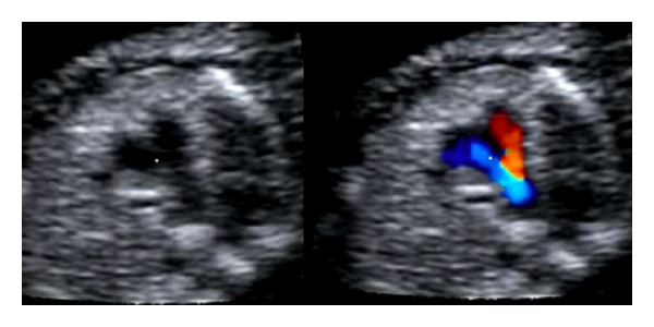
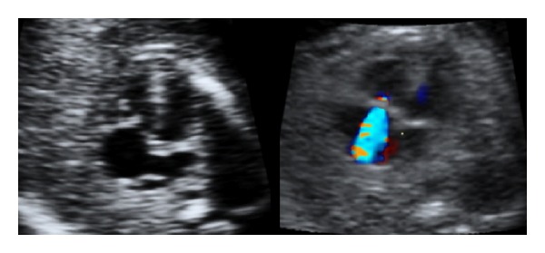
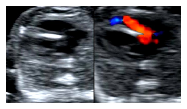
Objectives. To investigate the detection rate of major fetal heart defects in a low-risk population implementing routine use of color Doppler. Material and Methods. In a prospective observational study, all women undergoing fetal heart scanning (including 6781 routine examinations in the second trimester) during a three-year period were included. First a gray-scale scanning was performed including assessment of the four-chamber view and the great vessels. Thereafter three cross-sectional planes through the fetal thorax were assessed with color Doppler. Results. Thirty-nine fetuses had major heart defects, and 26 (67%) were prenatally detected. In 9/26 (35%) of cases the main ultrasound finding was related to the use of color Doppler. The survival rate of live born children was 91%. Conclusions. Routine use of color Doppler in fetal heart scanning in a low-risk population may be helpful in the detection of major heart defects; however, still severe malformations were missed prenatally.
目的。调查在实施常规彩色多普勒检查的低风险人群中主要胎儿心脏缺陷的检出率。材料与方法。在一项前瞻性观察研究中,纳入了三年期间所有接受胎儿心脏扫描的女性(包括孕中期的6781例常规检查)。首先进行灰阶扫描,包括评估四腔心切面和大血管。此后,用彩色多普勒评估通过胎儿胸部的三个横断面。结果。39例胎儿有主要心脏缺陷,其中26例(67%)在产前被检测出。在26例中的9例(35%)病例中,主要超声发现与彩色多普勒的使用有关。活产儿的存活率为91%。结论。在低风险人群的胎儿心脏扫描中常规使用彩色多普勒可能有助于检测主要心脏缺陷;然而,仍有严重畸形在产前被漏诊。