• 文献检索
  • 文档翻译
  • 深度研究
  • 学术资讯
  • Suppr Zotero 插件Zotero 插件
  • 邀请有礼
  • 套餐&价格
  • 历史记录
应用&插件
Suppr Zotero 插件Zotero 插件浏览器插件Mac 客户端Windows 客户端微信小程序
定价
高级版会员购买积分包购买API积分包
服务
文献检索文档翻译深度研究API 文档MCP 服务
关于我们
关于 Suppr公司介绍联系我们用户协议隐私条款
关注我们

Suppr 超能文献

核心技术专利:CN118964589B侵权必究
粤ICP备2023148730 号-1Suppr @ 2026

文献检索

告别复杂PubMed语法,用中文像聊天一样搜索,搜遍4000万医学文献。AI智能推荐,让科研检索更轻松。

立即免费搜索

文件翻译

保留排版,准确专业,支持PDF/Word/PPT等文件格式,支持 12+语言互译。

免费翻译文档

深度研究

AI帮你快速写综述,25分钟生成高质量综述,智能提取关键信息,辅助科研写作。

立即免费体验

相似文献

1
Vision-based proximity detection in retinal surgery.视网膜手术中的基于视觉的接近检测。
IEEE Trans Biomed Eng. 2012 Aug;59(8):2291-301. doi: 10.1109/TBME.2012.2202903. Epub 2012 Jun 5.
2
Hybrid tracking and mosaicking for information augmentation in retinal surgery.用于视网膜手术中信息增强的混合跟踪与拼接
Med Image Comput Comput Assist Interv. 2012;15(Pt 1):397-404. doi: 10.1007/978-3-642-33415-3_49.
3
Four-Dimensional Microscope-Integrated Optical Coherence Tomography Guidance in a Model Eye Subretinal Surgery.四维度显微镜整合于模型眼视网膜下手术之光学同调断层扫描引导
Retina. 2019 Oct;39 Suppl 1(Suppl 1):S194-S198. doi: 10.1097/IAE.0000000000002518.
4
Unified detection and tracking of instruments during retinal microsurgery.视网膜微创手术中器械的统一检测和跟踪。
IEEE Trans Pattern Anal Mach Intell. 2013 May;35(5):1263-73. doi: 10.1109/TPAMI.2012.209.
5
Unified detection and tracking in retinal microsurgery.视网膜显微手术中的统一检测与跟踪
Med Image Comput Comput Assist Interv. 2011;14(Pt 1):1-8. doi: 10.1007/978-3-642-23623-5_1.
6
Instrument tracking via online learning in retinal microsurgery.视网膜显微手术中通过在线学习进行器械跟踪
Med Image Comput Comput Assist Interv. 2014;17(Pt 1):464-71. doi: 10.1007/978-3-319-10404-1_58.
7
Force sensing micro-forceps for robot assisted retinal surgery.用于机器人辅助视网膜手术的力传感微型镊子。
Annu Int Conf IEEE Eng Med Biol Soc. 2012;2012:1401-4. doi: 10.1109/EMBC.2012.6346201.
8
Haptic interface for robot-assisted ophthalmic surgery.用于机器人辅助眼科手术的触觉接口。
Annu Int Conf IEEE Eng Med Biol Soc. 2015 Aug;2015:4906-9. doi: 10.1109/EMBC.2015.7319492.
9
Design and development of a computer-assisted retinal laser surgery system.计算机辅助视网膜激光手术系统的设计与开发。
J Biomed Opt. 2006 Jul-Aug;11(4):041127. doi: 10.1117/1.2342465.
10
CRF-Based Model for Instrument Detection and Pose Estimation in Retinal Microsurgery.基于条件随机场的视网膜显微手术器械检测与姿态估计模型
Comput Math Methods Med. 2016;2016:1067509. doi: 10.1155/2016/1067509. Epub 2016 Oct 27.

引用本文的文献

1
Fourier Lightfield Multiview Stereoscope for Large Field-of-View 3D Imaging in Microsurgical Settings.用于显微手术环境中大视野三维成像的傅里叶光场多视图立体显微镜
Adv Photonics Nexus. 2025 Jun;4(4). doi: 10.1117/1.apn.4.4.046008. Epub 2025 Jun 30.
2
Microsurgery Robots: Applications, Design, and Development.微创手术机器人:应用、设计与开发。
Sensors (Basel). 2023 Oct 16;23(20):8503. doi: 10.3390/s23208503.
3
Correction of Rotational Eccentricity Based on Model and Microvision in the Wire-Traction Micromanipulation System.基于模型与显微视觉的线牵引微操作系统中旋转偏心校正
Micromachines (Basel). 2023 Apr 28;14(5):963. doi: 10.3390/mi14050963.
4
Towards Autonomous Eye Surgery by Combining Deep Imitation Learning with Optimal Control.通过深度模仿学习与最优控制相结合实现自主眼部手术
Proc Mach Learn Res. 2021;155:2347-2358.
5
Autonomously Navigating a Surgical Tool Inside the Eye by Learning from Demonstration.通过示范学习实现眼内手术工具的自主导航。
IEEE Int Conf Robot Autom. 2020 May;2020. doi: 10.1109/icra40945.2020.9196537. Epub 2020 Sep 15.
6
Orthogonality Measurement of Three-Axis Motion Trajectories for Micromanipulation Robot Systems.微操作机器人系统三轴运动轨迹的正交性测量
Micromachines (Basel). 2021 Mar 23;12(3):344. doi: 10.3390/mi12030344.
7
Vision-Based Surgical Tool Pose Estimation for the da Vinci Robotic Surgical System.用于达芬奇机器人手术系统的基于视觉的手术工具位姿估计
Rep U S. 2018 Oct;2018:1298-1305. doi: 10.1109/IROS.2018.8594471. Epub 2019 Jan 7.
8
Techniques for robot-aided intraocular surgery using monocular vision.使用单眼视觉的机器人辅助眼内手术技术。
Int J Rob Res. 2018 Jul;37(8):931-952. doi: 10.1177/0278364918778352. Epub 2018 Jul 13.
9
Toward Monocular Camera-Guided Retinal Vein Cannulation with an Actively Stabilized Handheld Robot.迈向基于单目相机引导、由主动稳定的手持机器人进行的视网膜静脉插管技术。
IEEE Int Conf Robot Autom. 2017 May-Jun;2017:2951-2956. doi: 10.1109/ICRA.2017.7989341. Epub 2017 Jul 24.
10
Surgical data science: The new knowledge domain.外科数据科学:新的知识领域。
Innov Surg Sci. 2017 Apr;2(3):109-121. doi: 10.1515/iss-2017-0004. Epub 2017 Apr 20.

本文引用的文献

1
Unified detection and tracking in retinal microsurgery.视网膜显微手术中的统一检测与跟踪
Med Image Comput Comput Assist Interv. 2011;14(Pt 1):1-8. doi: 10.1007/978-3-642-23623-5_1.
2
Retinal vessel cannulation with an image-guided handheld robot.使用图像引导的手持机器人进行视网膜血管插管。
Annu Int Conf IEEE Eng Med Biol Soc. 2010;2010:5420-3. doi: 10.1109/IEMBS.2010.5626493.
3
Adaptive multispectral illumination for retinal microsurgery.用于视网膜显微手术的自适应多光谱照明
Med Image Comput Comput Assist Interv. 2010;13(Pt 3):465-72. doi: 10.1007/978-3-642-15711-0_58.
4
Tracking of irregular graphical structures for tissue deformation recovery in minimally invasive surgery.用于微创手术中组织变形恢复的不规则图形结构跟踪
Med Image Comput Comput Assist Interv. 2010;13(Pt 3):261-8. doi: 10.1007/978-3-642-15711-0_33.
5
Real-time stereo reconstruction in robotically assisted minimally invasive surgery.机器人辅助微创手术中的实时立体重建
Med Image Comput Comput Assist Interv. 2010;13(Pt 1):275-82. doi: 10.1007/978-3-642-15705-9_34.
6
Single fiber optical coherence tomography microsurgical instruments for computer and robot-assisted retinal surgery.用于计算机和机器人辅助视网膜手术的单光纤光学相干断层扫描显微手术器械。
Med Image Comput Comput Assist Interv. 2009;12(Pt 1):108-15. doi: 10.1007/978-3-642-04268-3_14.
7
A sub-millimetric, 0.25 mN resolution fully integrated fiber-optic force-sensing tool for retinal microsurgery.一种用于视网膜微创手术的亚毫米、0.25 毫牛分辨率的全集成光纤测力工具。
Int J Comput Assist Radiol Surg. 2009 Jun;4(4):383-90. doi: 10.1007/s11548-009-0301-6. Epub 2009 Apr 15.
8
A new multi-modal similarity measure for fast gradient-based 2D-3D image registration.一种用于基于快速梯度的2D-3D图像配准的新型多模态相似性度量。
Annu Int Conf IEEE Eng Med Biol Soc. 2009;2009:5821-4. doi: 10.1109/IEMBS.2009.5335172.
9
Groupwise geometric and photometric direct image registration.逐组几何和光度直接图像配准。
IEEE Trans Pattern Anal Mach Intell. 2008 Dec;30(12):2098-108. doi: 10.1109/TPAMI.2008.22.
10
Tracking endoscopic instruments without a localizer: a shape-analysis-based approach.无需定位器的内镜器械跟踪:一种基于形状分析的方法。
Comput Aided Surg. 2007 Jan;12(1):35-42. doi: 10.3109/10929080701210782.

视网膜手术中的基于视觉的接近检测。

Vision-based proximity detection in retinal surgery.

机构信息

Laboratory of Computational Sensing and Robotics, The Johns Hopkins University, Baltimore, MD 21218, USA.

出版信息

IEEE Trans Biomed Eng. 2012 Aug;59(8):2291-301. doi: 10.1109/TBME.2012.2202903. Epub 2012 Jun 5.

DOI:10.1109/TBME.2012.2202903
PMID:22692869
原文链接:https://pmc.ncbi.nlm.nih.gov/articles/PMC3467359/
Abstract

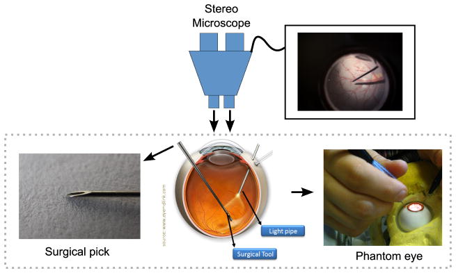
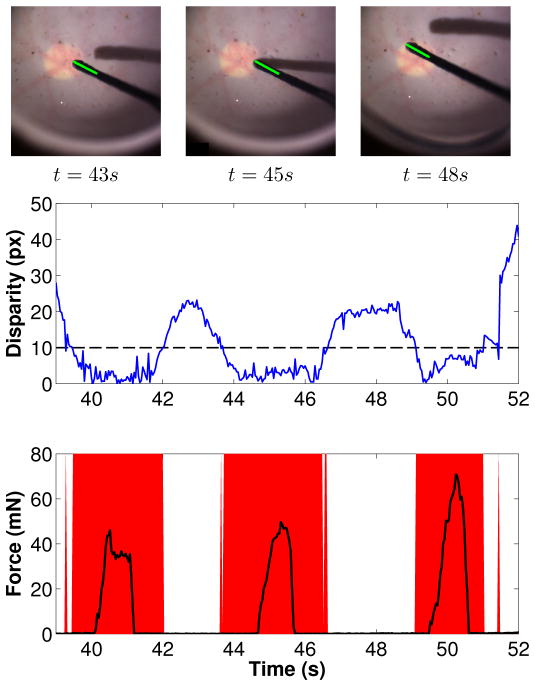
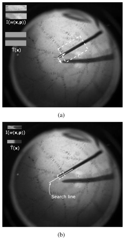
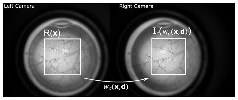
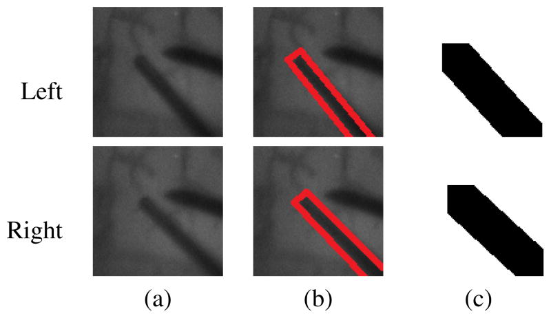
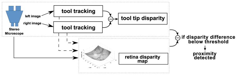
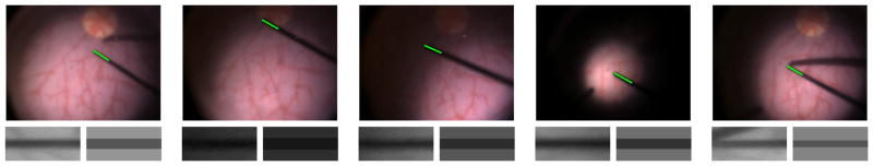
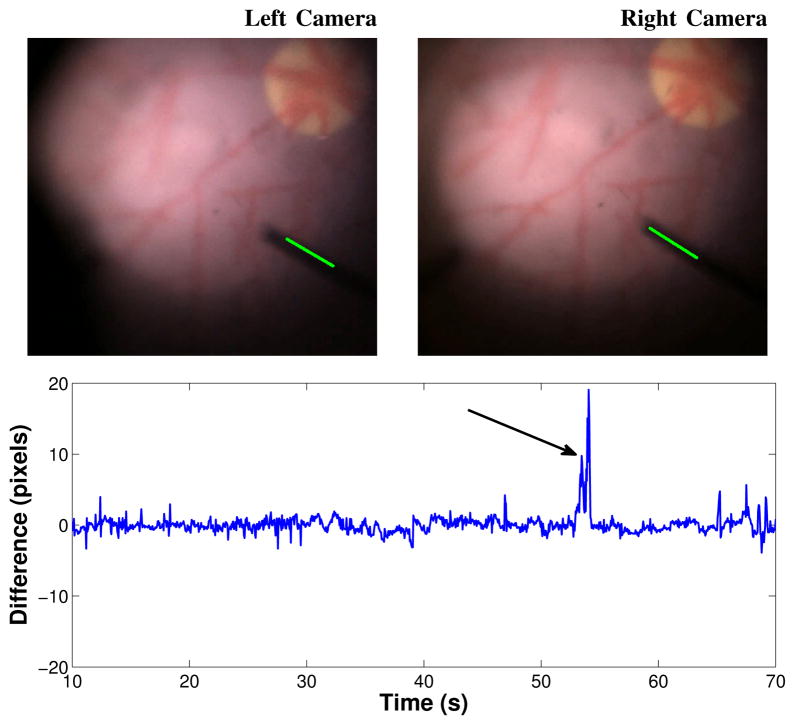
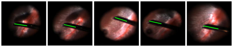
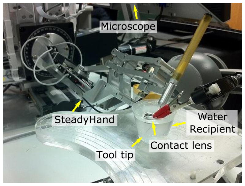
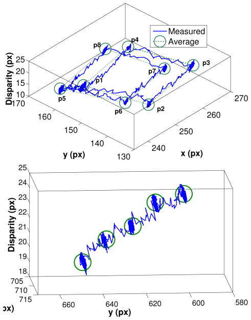
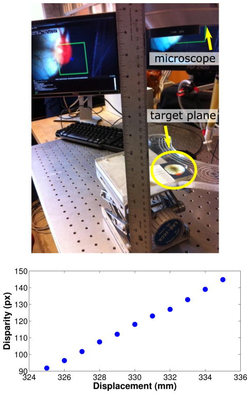
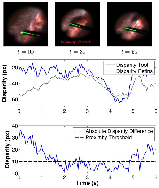
In retinal surgery, surgeons face difficulties such as indirect visualization of surgical targets, physiological tremor, and lack of tactile feedback, which increase the risk of retinal damage caused by incorrect surgical gestures. In this context, intraocular proximity sensing has the potential to overcome current technical limitations and increase surgical safety. In this paper, we present a system for detecting unintentional collisions between surgical tools and the retina using the visual feedback provided by the opthalmic stereo microscope. Using stereo images, proximity between surgical tools and the retinal surface can be detected when their relative stereo disparity is small. For this purpose, we developed a system comprised of two modules. The first is a module for tracking the surgical tool position on both stereo images. The second is a disparity tracking module for estimating a stereo disparity map of the retinal surface. Both modules were specially tailored for coping with the challenging visualization conditions in retinal surgery. The potential clinical value of the proposed method is demonstrated by extensive testing using a silicon phantom eye and recorded rabbit in vivo data.

摘要

在视网膜手术中,外科医生面临间接可视化手术目标、生理震颤和缺乏触觉反馈等困难,这些都增加了因手术手势不当而导致视网膜损伤的风险。在这种情况下,眼内接近感应有可能克服当前的技术限制,提高手术安全性。在本文中,我们提出了一种使用眼科立体显微镜提供的视觉反馈来检测手术工具与视网膜之间意外碰撞的系统。使用立体图像,当手术工具和视网膜表面之间的相对立体视差较小时,可以检测到它们之间的接近程度。为此,我们开发了一个由两个模块组成的系统。第一个模块是用于在两幅立体图像上跟踪手术工具位置的模块。第二个模块是用于估计视网膜表面立体视差图的视差跟踪模块。这两个模块都是专门为应对视网膜手术中具有挑战性的可视化条件而设计的。通过使用硅质人工眼球和记录的兔子体内数据进行广泛测试,证明了所提出方法的潜在临床价值。

https://cdn.ncbi.nlm.nih.gov/pmc/blobs/fe21/3467359/deda7da70821/nihms386690f15.jpg
https://cdn.ncbi.nlm.nih.gov/pmc/blobs/fe21/3467359/356198c01f26/nihms386690f1.jpg
https://cdn.ncbi.nlm.nih.gov/pmc/blobs/fe21/3467359/6dfcb94d10bf/nihms386690f2.jpg
https://cdn.ncbi.nlm.nih.gov/pmc/blobs/fe21/3467359/65f57673744c/nihms386690f3.jpg
https://cdn.ncbi.nlm.nih.gov/pmc/blobs/fe21/3467359/8706d2428e82/nihms386690f4.jpg
https://cdn.ncbi.nlm.nih.gov/pmc/blobs/fe21/3467359/1f41ecac20c5/nihms386690f5.jpg
https://cdn.ncbi.nlm.nih.gov/pmc/blobs/fe21/3467359/71e308ef0948/nihms386690f6.jpg
https://cdn.ncbi.nlm.nih.gov/pmc/blobs/fe21/3467359/610af2df2edb/nihms386690f7.jpg
https://cdn.ncbi.nlm.nih.gov/pmc/blobs/fe21/3467359/732d58875dfa/nihms386690f8.jpg
https://cdn.ncbi.nlm.nih.gov/pmc/blobs/fe21/3467359/9c1f0f990fca/nihms386690f9.jpg
https://cdn.ncbi.nlm.nih.gov/pmc/blobs/fe21/3467359/77f4254830c7/nihms386690f10.jpg
https://cdn.ncbi.nlm.nih.gov/pmc/blobs/fe21/3467359/8925bd26e9f5/nihms386690f11.jpg
https://cdn.ncbi.nlm.nih.gov/pmc/blobs/fe21/3467359/1f98bcfab1cd/nihms386690f12.jpg
https://cdn.ncbi.nlm.nih.gov/pmc/blobs/fe21/3467359/f23e6c7fddc5/nihms386690f13.jpg
https://cdn.ncbi.nlm.nih.gov/pmc/blobs/fe21/3467359/70f3890cd7ea/nihms386690f14.jpg
https://cdn.ncbi.nlm.nih.gov/pmc/blobs/fe21/3467359/deda7da70821/nihms386690f15.jpg