• 文献检索
  • 文档翻译
  • 深度研究
  • 学术资讯
  • Suppr Zotero 插件Zotero 插件
  • 邀请有礼
  • 套餐&价格
  • 历史记录
应用&插件
Suppr Zotero 插件Zotero 插件浏览器插件Mac 客户端Windows 客户端微信小程序
定价
高级版会员购买积分包购买API积分包
服务
文献检索文档翻译深度研究API 文档MCP 服务
关于我们
关于 Suppr公司介绍联系我们用户协议隐私条款
关注我们

Suppr 超能文献

核心技术专利:CN118964589B侵权必究
粤ICP备2023148730 号-1Suppr @ 2026

文献检索

告别复杂PubMed语法,用中文像聊天一样搜索,搜遍4000万医学文献。AI智能推荐,让科研检索更轻松。

立即免费搜索

文件翻译

保留排版,准确专业,支持PDF/Word/PPT等文件格式,支持 12+语言互译。

免费翻译文档

深度研究

AI帮你快速写综述,25分钟生成高质量综述,智能提取关键信息,辅助科研写作。

立即免费体验

基于分层补丁的稀疏表示——一种用于提高 4D-CT 肺部数据分辨率的新方法。

Hierarchical patch-based sparse representation--a new approach for resolution enhancement of 4D-CT lung data.

出版信息

IEEE Trans Med Imaging. 2012 Nov;31(11):1993-2005. doi: 10.1109/TMI.2012.2202245. Epub 2012 Jun 6.

DOI:10.1109/TMI.2012.2202245
PMID:22692897
原文链接:https://pmc.ncbi.nlm.nih.gov/articles/PMC11166181/
Abstract

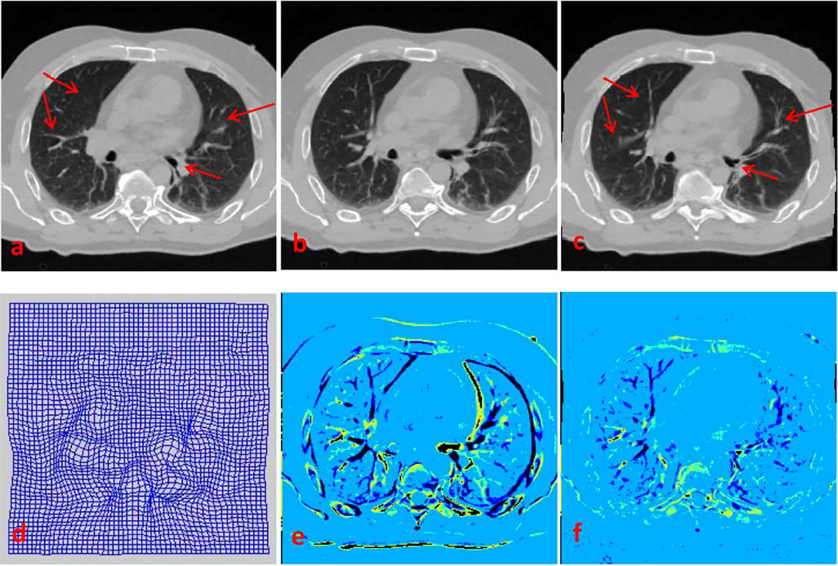
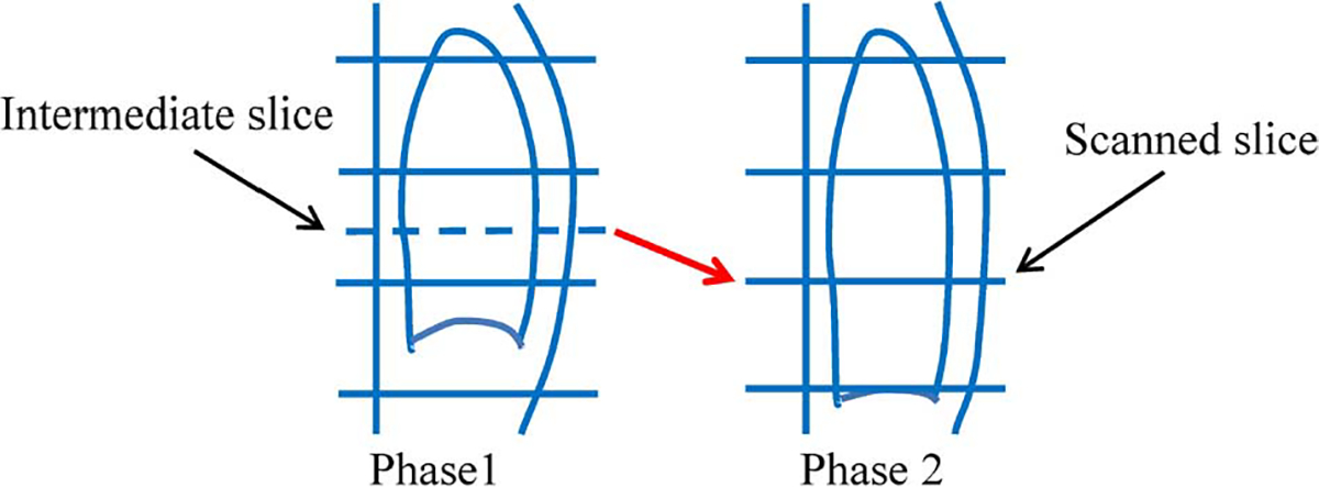
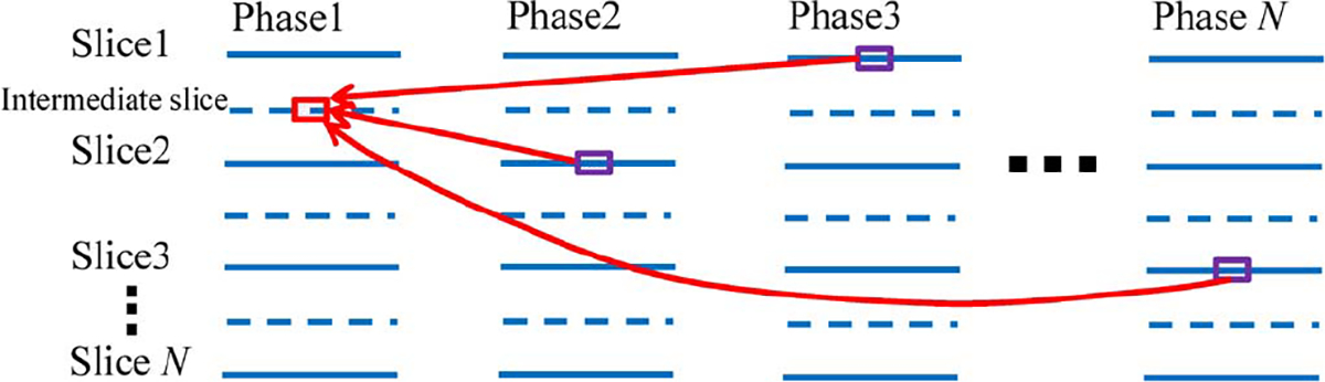
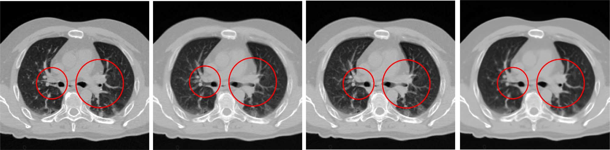
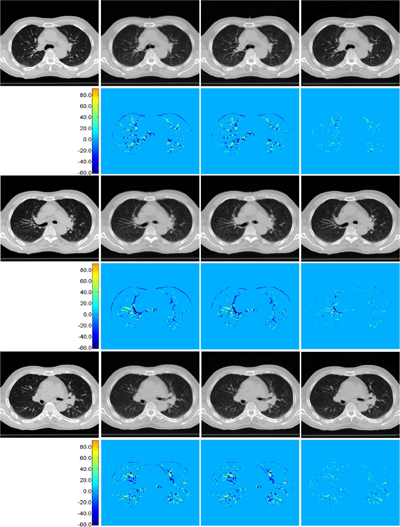
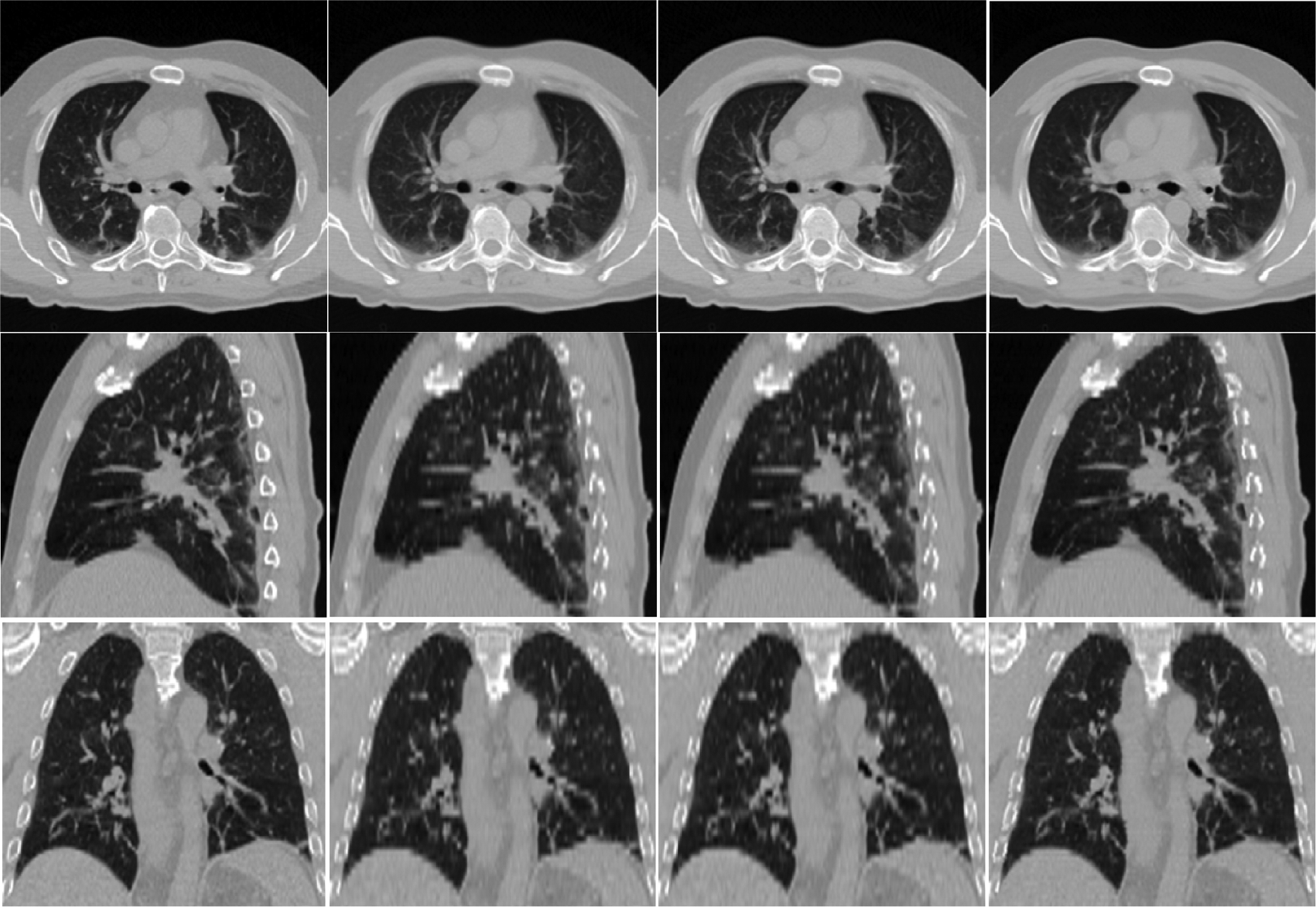
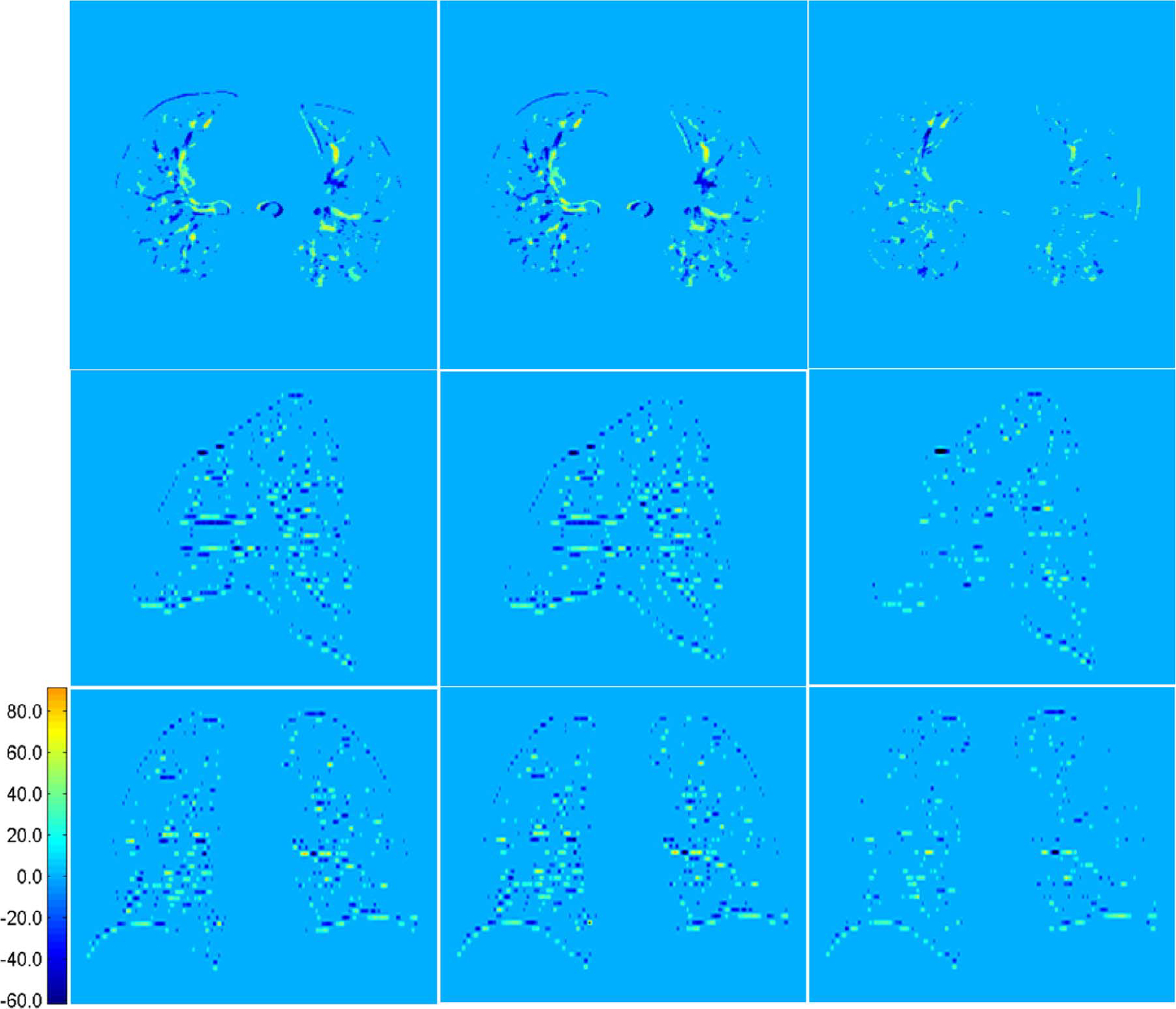
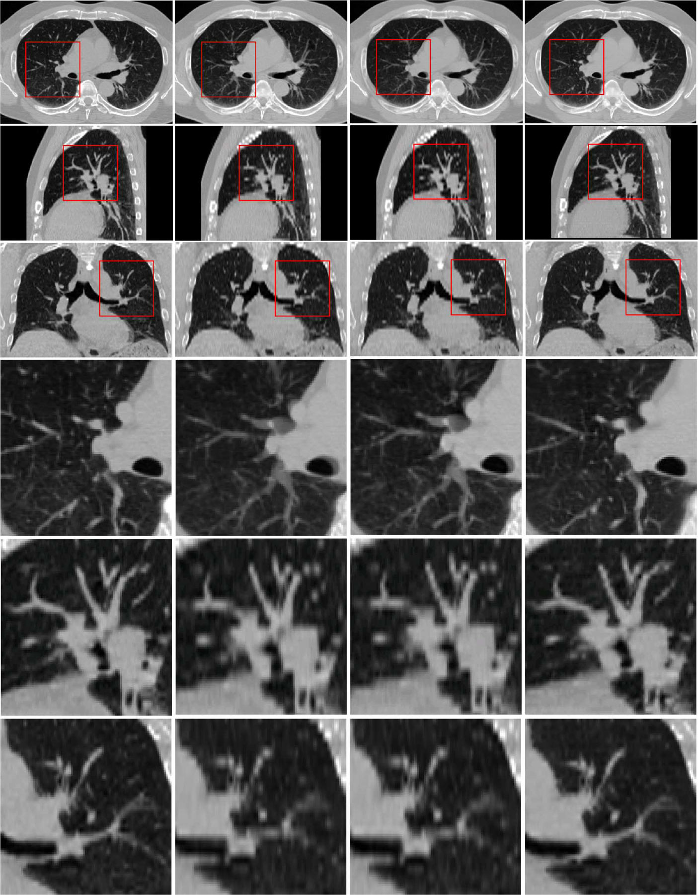
4D-CT plays an important role in lung cancer treatment because of its capability in providing a comprehensive characterization of respiratory motion for high-precision radiation therapy. However, due to the inherent high-dose exposure associated with CT, dense sampling along superior-inferior direction is often not practical, thus resulting in an inter-slice thickness that is much greater than in-plane voxel resolutions. As a consequence, artifacts such as lung vessel discontinuity and partial volume effects are often observed in 4D-CT images, which may mislead dose administration in radiation therapy. In this paper, we present a novel patch-based technique for resolution enhancement of 4D-CT images along the superior-inferior direction. Our working premise is that anatomical information that is missing in one particular phase can be recovered from other phases. Based on this assumption, we employ a hierarchical patch-based sparse representation mechanism to enhance the superior-inferior resolution of 4D-CT by reconstructing additional intermediate CT slices. Specifically, for each spatial location on an intermediate CT slice that we intend to reconstruct, we first agglomerate a dictionary of patches from images of all other phases in the 4D-CT. We then employ a sparse combination of patches from this dictionary, with guidance from neighboring (upper and lower) slices, to reconstruct a series of patches, which we progressively refine in a hierarchical fashion to reconstruct the final intermediate slices with significantly enhanced anatomical details. Our method was extensively evaluated using a public dataset. In all experiments, our method outperforms the conventional linear and cubic-spline interpolation methods in preserving image details and also in suppressing misleading artifacts, indicating that our proposed method can potentially be applied to better image-guided radiation therapy of lung cancer in the future.

摘要

4D-CT 在肺癌治疗中起着重要作用,因为它能够全面描述呼吸运动,从而实现高精度放射治疗。然而,由于 CT 固有的高剂量暴露,沿上下方向进行密集采样通常是不切实际的,因此导致切片间厚度远大于平面体素分辨率。结果,4D-CT 图像中经常观察到肺部血管不连续和部分容积效应等伪影,这可能会导致放射治疗中的剂量给药错误。在本文中,我们提出了一种新颖的基于补丁的技术,用于增强 4D-CT 图像在上下方向上的分辨率。我们的工作前提是,在特定相位中丢失的解剖信息可以从其他相位中恢复。基于此假设,我们采用分层基于补丁的稀疏表示机制,通过重建额外的中间 CT 切片来增强 4D-CT 的上下分辨率。具体来说,对于我们要重建的中间 CT 切片上的每个空间位置,我们首先从 4D-CT 中的所有其他相位的图像中聚集补丁字典。然后,我们使用来自该字典的补丁的稀疏组合,并通过来自上下相邻切片的指导,重建一系列补丁,我们以分层的方式逐步细化这些补丁,以重建具有显著增强解剖细节的最终中间切片。我们的方法在一个公共数据集上进行了广泛评估。在所有实验中,我们的方法在保留图像细节和抑制误导性伪影方面都优于传统的线性和三次样条插值方法,这表明我们提出的方法将来可能会应用于更好的肺癌图像引导放射治疗。

https://cdn.ncbi.nlm.nih.gov/pmc/blobs/25c7/11166181/a351a74f0506/nihms-1996512-f0017.jpg
https://cdn.ncbi.nlm.nih.gov/pmc/blobs/25c7/11166181/f2232317702f/nihms-1996512-f0001.jpg
https://cdn.ncbi.nlm.nih.gov/pmc/blobs/25c7/11166181/05a952a17468/nihms-1996512-f0002.jpg
https://cdn.ncbi.nlm.nih.gov/pmc/blobs/25c7/11166181/e6afaa90ebc7/nihms-1996512-f0003.jpg
https://cdn.ncbi.nlm.nih.gov/pmc/blobs/25c7/11166181/a5ee5cfe7fe8/nihms-1996512-f0004.jpg
https://cdn.ncbi.nlm.nih.gov/pmc/blobs/25c7/11166181/44ecbf40230a/nihms-1996512-f0005.jpg
https://cdn.ncbi.nlm.nih.gov/pmc/blobs/25c7/11166181/2b25b9970518/nihms-1996512-f0006.jpg
https://cdn.ncbi.nlm.nih.gov/pmc/blobs/25c7/11166181/abdcacd2c323/nihms-1996512-f0007.jpg
https://cdn.ncbi.nlm.nih.gov/pmc/blobs/25c7/11166181/6dc1f881ebe1/nihms-1996512-f0008.jpg
https://cdn.ncbi.nlm.nih.gov/pmc/blobs/25c7/11166181/4cf9451e2546/nihms-1996512-f0009.jpg
https://cdn.ncbi.nlm.nih.gov/pmc/blobs/25c7/11166181/fe1b7dbfcd22/nihms-1996512-f0010.jpg
https://cdn.ncbi.nlm.nih.gov/pmc/blobs/25c7/11166181/881f1a9da505/nihms-1996512-f0011.jpg
https://cdn.ncbi.nlm.nih.gov/pmc/blobs/25c7/11166181/658ce23885d3/nihms-1996512-f0012.jpg
https://cdn.ncbi.nlm.nih.gov/pmc/blobs/25c7/11166181/5fa2526f66c2/nihms-1996512-f0013.jpg
https://cdn.ncbi.nlm.nih.gov/pmc/blobs/25c7/11166181/565d74244be1/nihms-1996512-f0014.jpg
https://cdn.ncbi.nlm.nih.gov/pmc/blobs/25c7/11166181/f309e0a84824/nihms-1996512-f0015.jpg
https://cdn.ncbi.nlm.nih.gov/pmc/blobs/25c7/11166181/702c5e7b04a3/nihms-1996512-f0016.jpg
https://cdn.ncbi.nlm.nih.gov/pmc/blobs/25c7/11166181/a351a74f0506/nihms-1996512-f0017.jpg

相似文献

1
Hierarchical patch-based sparse representation--a new approach for resolution enhancement of 4D-CT lung data.基于分层补丁的稀疏表示——一种用于提高 4D-CT 肺部数据分辨率的新方法。
IEEE Trans Med Imaging. 2012 Nov;31(11):1993-2005. doi: 10.1109/TMI.2012.2202245. Epub 2012 Jun 6.
2
Resolution enhancement of lung 4D-CT via group-sparsity.基于群组稀疏的肺部 4D-CT 分辨率增强。
Med Phys. 2013 Dec;40(12):121717. doi: 10.1118/1.4829501.
3
Resolution enhancement of lung 4D-CT data using multiscale interphase iterative nonlocal means.使用多尺度相间迭代非局部均值提高肺部 4D-CT 数据的分辨率。
Med Phys. 2013 May;40(5):051916. doi: 10.1118/1.4802747.
4
Improving image-guided radiation therapy of lung cancer by reconstructing 4D-CT from a single free-breathing 3D-CT on the treatment day.在治疗当天,通过从单次自由呼吸 3D-CT 重建 4D-CT,改善肺癌的图像引导放射治疗。
Med Phys. 2012 Dec;39(12):7694-709. doi: 10.1118/1.4768226.
5
Harnessing group-sparsity regularization for resolution enhancement of lung 4D-CT.利用组稀疏正则化提高肺部4D-CT分辨率
Med Image Comput Comput Assist Interv. 2013;16(Pt 3):139-46. doi: 10.1007/978-3-642-40760-4_18.
6
Non-local means resolution enhancement of lung 4D-CT data.肺部4D-CT数据的非局部均值分辨率增强
Med Image Comput Comput Assist Interv. 2012;15(Pt 1):214-22. doi: 10.1007/978-3-642-33415-3_27.
7
Registration based super-resolution reconstruction for lung 4D-CT.基于配准的肺部4D-CT超分辨率重建
Annu Int Conf IEEE Eng Med Biol Soc. 2014;2014:2444-7. doi: 10.1109/EMBC.2014.6944116.
8
Super-resolution reconstruction of 4D-CT lung data via patch-based low-rank matrix reconstruction.基于块的低秩矩阵重建实现4D-CT肺部数据的超分辨率重建
Phys Med Biol. 2017 Oct 3;62(20):7925-7937. doi: 10.1088/1361-6560/aa8a48.
9
Super-resolution reconstruction for 4D computed tomography of the lung via the projections onto convex sets approach.通过凸集投影法实现肺部四维计算机断层扫描的超分辨率重建。
Med Phys. 2014 Nov;41(11):111917. doi: 10.1118/1.4899185.
10
Modeling respiratory motion for reducing motion artifacts in 4D CT images.建立呼吸运动模型以减少 4D CT 图像中的运动伪影。
Med Phys. 2013 Apr;40(4):041716. doi: 10.1118/1.4795133.

引用本文的文献

1
Deep learning-assisted terahertz intelligent detection and identification of cancer tissue.深度学习辅助的太赫兹癌症组织智能检测与识别
Fundam Res. 2025 Mar 26;5(4):1831-1842. doi: 10.1016/j.fmre.2025.03.013. eCollection 2025 Jul.
2
Generative adversarial networks with adaptive normalization for synthesizing T2-weighted magnetic resonance images from diffusion-weighted images.用于从扩散加权图像合成T2加权磁共振图像的具有自适应归一化的生成对抗网络。
Front Neurosci. 2022 Nov 14;16:1058487. doi: 10.3389/fnins.2022.1058487. eCollection 2022.
3
Synthesized 7T MRI from 3T MRI via deep learning in spatial and wavelet domains.

本文引用的文献

1
Super-resolution in respiratory synchronized positron emission tomography.呼吸门控正电子发射断层扫描中的超分辨率。
IEEE Trans Med Imaging. 2012 Feb;31(2):438-48. doi: 10.1109/TMI.2011.2171358. Epub 2011 Oct 13.
2
Low-dose 4DCT reconstruction via temporal nonlocal means.基于时域非局部均值的低剂量 4DCT 重建。
Med Phys. 2011 Mar;38(3):1359-65. doi: 10.1118/1.3547724.
3
MRI RESOLUTION ENHANCEMENT USING TOTAL VARIATION REGULARIZATION.使用全变差正则化的磁共振成像分辨率增强
基于深度学习的空间域和小波域的 3T MRI 向 7T MRI 的合成。
Med Image Anal. 2020 May;62:101663. doi: 10.1016/j.media.2020.101663. Epub 2020 Feb 19.
4
Dual-domain convolutional neural networks for improving structural information in 3 T MRI.双域卷积神经网络提高 3T MRI 结构信息。
Magn Reson Imaging. 2019 Dec;64:90-100. doi: 10.1016/j.mri.2019.05.023. Epub 2019 Jun 5.
5
Brain lesion segmentation through image synthesis and outlier detection.通过图像合成和异常值检测进行脑损伤分割。
Neuroimage Clin. 2017 Sep 8;16:643-658. doi: 10.1016/j.nicl.2017.09.003. eCollection 2017.
6
7T-guided super-resolution of 3T MRI.7T 引导下的 3T MRI 超分辨率
Med Phys. 2017 May;44(5):1661-1677. doi: 10.1002/mp.12132. Epub 2017 Apr 22.
7
Segmentation Based Sparse Reconstruction of Optical Coherence Tomography Images.基于分割的光学相干断层扫描图像稀疏重建
IEEE Trans Med Imaging. 2017 Feb;36(2):407-421. doi: 10.1109/TMI.2016.2611503. Epub 2016 Sep 20.
8
Reconstruction of 7T-Like Images From 3T MRI.从3T磁共振成像重建类似7T的图像。
IEEE Trans Med Imaging. 2016 Sep;35(9):2085-97. doi: 10.1109/TMI.2016.2549918. Epub 2016 Apr 1.
9
Dictionary-Driven Ischemia Detection From Cardiac Phase-Resolved Myocardial BOLD MRI at Rest.基于字典驱动的静息状态下心脏相位分辨心肌血氧水平依赖性功能磁共振成像的缺血检测
IEEE Trans Med Imaging. 2016 Jan;35(1):282-93. doi: 10.1109/TMI.2015.2470075. Epub 2015 Aug 19.
10
3-D Adaptive Sparsity Based Image Compression With Applications to Optical Coherence Tomography.基于三维自适应稀疏性的图像压缩及其在光学相干断层扫描中的应用
IEEE Trans Med Imaging. 2015 Jun;34(6):1306-20. doi: 10.1109/TMI.2014.2387336. Epub 2015 Jan 1.
Proc IEEE Int Symp Biomed Imaging. 2009 Aug 7;2009:161-164. doi: 10.1109/ISBI.2009.5193008.
4
MR image reconstruction from highly undersampled k-space data by dictionary learning.基于字典学习的欠采样 k 空间数据磁共振图像重建。
IEEE Trans Med Imaging. 2011 May;30(5):1028-41. doi: 10.1109/TMI.2010.2090538. Epub 2010 Nov 1.
5
A non-local approach for image super-resolution using intermodality priors.基于跨模态先验的非局部图像超分辨率方法。
Med Image Anal. 2010 Aug;14(4):594-605. doi: 10.1016/j.media.2010.04.005. Epub 2010 May 6.
6
Intensity-based image registration by minimizing residual complexity.基于残差复杂度最小化的强度图像配准。
IEEE Trans Med Imaging. 2010 Nov;29(11):1882-91. doi: 10.1109/TMI.2010.2053043. Epub 2010 Jun 17.
7
Robust super-resolution volume reconstruction from slice acquisitions: application to fetal brain MRI.从切片采集进行稳健的超分辨率体积重建:在胎儿脑 MRI 中的应用。
IEEE Trans Med Imaging. 2010 Oct;29(10):1739-58. doi: 10.1109/TMI.2010.2051680. Epub 2010 Jun 7.
8
Image super-resolution via sparse representation.基于稀疏表示的图像超分辨率重建。
IEEE Trans Image Process. 2010 Nov;19(11):2861-73. doi: 10.1109/TIP.2010.2050625. Epub 2010 May 18.
9
Image inpainting by patch propagation using patch sparsity.基于补丁稀疏性的补丁传播图像修复。
IEEE Trans Image Process. 2010 May;19(5):1153-65. doi: 10.1109/TIP.2010.2042098. Epub 2010 Feb 2.
10
The use of 4DCT to reduce lung dose: a dosimetric analysis.使用4DCT降低肺部剂量:剂量学分析。
Med Dosim. 2009 Winter;34(4):273-8. doi: 10.1016/j.meddos.2008.11.005. Epub 2009 Jan 1.