• 文献检索
  • 文档翻译
  • 深度研究
  • 学术资讯
  • Suppr Zotero 插件Zotero 插件
  • 邀请有礼
  • 套餐&价格
  • 历史记录
应用&插件
Suppr Zotero 插件Zotero 插件浏览器插件Mac 客户端Windows 客户端微信小程序
定价
高级版会员购买积分包购买API积分包
服务
文献检索文档翻译深度研究API 文档MCP 服务
关于我们
关于 Suppr公司介绍联系我们用户协议隐私条款
关注我们

Suppr 超能文献

核心技术专利:CN118964589B侵权必究
粤ICP备2023148730 号-1Suppr @ 2026

文献检索

告别复杂PubMed语法,用中文像聊天一样搜索,搜遍4000万医学文献。AI智能推荐,让科研检索更轻松。

立即免费搜索

文件翻译

保留排版,准确专业,支持PDF/Word/PPT等文件格式,支持 12+语言互译。

免费翻译文档

深度研究

AI帮你快速写综述,25分钟生成高质量综述,智能提取关键信息,辅助科研写作。

立即免费体验

使用动态容积 CT 进行灌注成像时,自由呼吸与屏气的比较。

Comparison of free breathing versus breath-hold in perfusion imaging using dynamic volume CT.

机构信息

Department of Medical Imaging, University of Toronto, Toronto General Hospital, NCSB 1C547, 585 University Avenue, Toronto, Ontario, M5G 2N2, Canada,

出版信息

Insights Imaging. 2012 Aug;3(4):323-8. doi: 10.1007/s13244-012-0169-9. Epub 2012 May 1.

DOI:10.1007/s13244-012-0169-9
PMID:22695948
原文链接:https://pmc.ncbi.nlm.nih.gov/articles/PMC3481081/
Abstract

OBJECTIVES

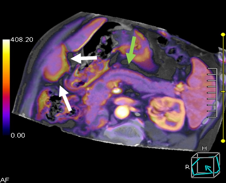
To compare two scanning protocols (free breathing versus breath-hold) for perfusion imaging using dynamic volume computed tomography (CT) and to evaluate their effects on image registration.

MATERIAL AND METHODS

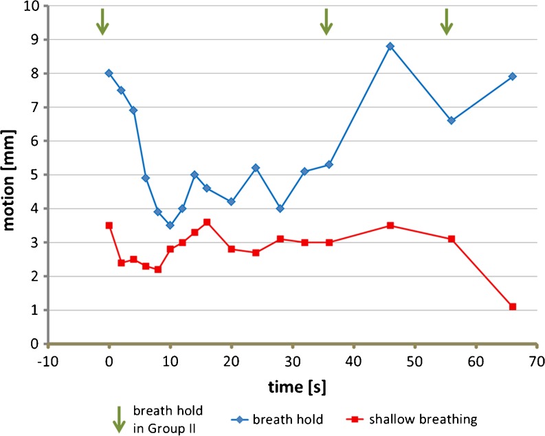
Forty patients underwent dynamic volume CT for pancreatic perfusion analysis and were randomly assigned to either a shallow-breathing (I) or breath-hold (II) group. Both dynamic CT protocols consisted of 17 low-dose volumetric scans. Rigid image registration was performed by using the volume with highest aortic attenuation as reference. All other volumes were visually matched with the pancreatic lesion serving as the volumetric region of interest. The overall demand for post-processing per patient was calculated as the median of three-dimensional vector lengths of all volumes in relation to the relative patient origin. The number of volumes not requiring registration was recorded per group.

RESULTS

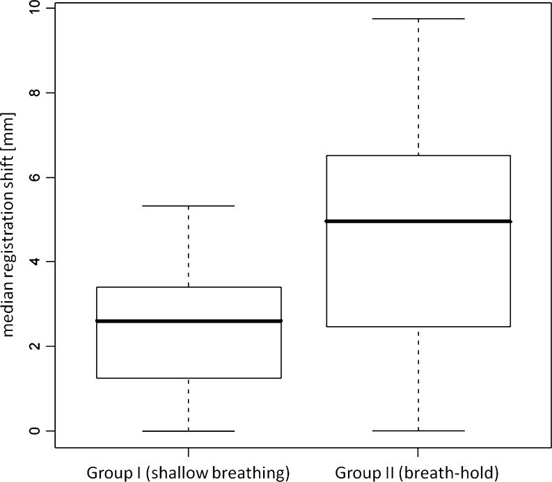
Registration mismatch for groups I and II was 2.61 mm (SD, 1.57) and 4.95 mm (SD, 2.71), respectively (P < 0.005). Twenty-eight volumes in group I (8.2%) and 47 volumes in group II (14.1%) did not require manual registration (P = 0.014).

CONCLUSION

Shallow breathing during dynamic volume CT scanning reduces the overall demand for motion correction and thus may be beneficial in perfusion imaging of the pancreas

MAIN MESSAGES

• Shallow breathing during perfusion CT scanning reduces the overall demand for motion correction. • Shallow breathing may be beneficial in perfusion imaging of the pancreas. • Image registration is crucial for CT perfusion imaging.

摘要

目的

比较两种扫描方案(自由呼吸与屏气)在使用动态容积 CT(CT)进行灌注成像时的效果,并评估其对图像配准的影响。

材料与方法

40 例患者行胰腺灌注动态容积 CT 检查,随机分为浅呼吸组(I 组)和屏气组(II 组)。两种动态 CT 方案均由 17 个低剂量容积扫描组成。采用主动脉衰减值最高的容积作为参考进行刚性图像配准。所有其他容积均与胰腺病变进行视觉匹配,作为容积感兴趣区。每位患者的后处理总需求计算为与患者原点相关的所有容积的三维向量长度的中位数。记录每组无需配准的容积数。

结果

I 组和 II 组的配准不匹配分别为 2.61mm(SD,1.57)和 4.95mm(SD,2.71)(P<0.005)。I 组有 28 个容积(8.2%),II 组有 47 个容积(14.1%)无需手动配准(P=0.014)。

结论

动态容积 CT 扫描时浅呼吸可减少运动校正的总体需求,因此可能对胰腺灌注成像有益。

主要信息

• 灌注 CT 扫描时浅呼吸可减少运动校正的总体需求。• 浅呼吸可能对胰腺灌注成像有益。• 图像配准对 CT 灌注成像至关重要。

https://cdn.ncbi.nlm.nih.gov/pmc/blobs/88d7/3481081/2e58189e0513/13244_2012_169_Fig3_HTML.jpg
https://cdn.ncbi.nlm.nih.gov/pmc/blobs/88d7/3481081/851b95f8e541/13244_2012_169_Fig1_HTML.jpg
https://cdn.ncbi.nlm.nih.gov/pmc/blobs/88d7/3481081/e9c476804612/13244_2012_169_Fig2_HTML.jpg
https://cdn.ncbi.nlm.nih.gov/pmc/blobs/88d7/3481081/2e58189e0513/13244_2012_169_Fig3_HTML.jpg
https://cdn.ncbi.nlm.nih.gov/pmc/blobs/88d7/3481081/851b95f8e541/13244_2012_169_Fig1_HTML.jpg
https://cdn.ncbi.nlm.nih.gov/pmc/blobs/88d7/3481081/e9c476804612/13244_2012_169_Fig2_HTML.jpg
https://cdn.ncbi.nlm.nih.gov/pmc/blobs/88d7/3481081/2e58189e0513/13244_2012_169_Fig3_HTML.jpg

相似文献

1
Comparison of free breathing versus breath-hold in perfusion imaging using dynamic volume CT.使用动态容积 CT 进行灌注成像时,自由呼吸与屏气的比较。
Insights Imaging. 2012 Aug;3(4):323-8. doi: 10.1007/s13244-012-0169-9. Epub 2012 May 1.
2
Real-time respiratory triggered SPECT myocardial perfusion imaging using CZT technology: impact of respiratory phase matching between SPECT and low-dose CT for attenuation correction.使用CZT技术的实时呼吸触发单光子发射计算机断层扫描心肌灌注成像:单光子发射计算机断层扫描与低剂量计算机断层扫描之间呼吸相位匹配对衰减校正的影响
Eur Heart J Cardiovasc Imaging. 2017 Jan;18(1):31-38. doi: 10.1093/ehjci/jew031. Epub 2016 Mar 16.
3
Internal target volume determined with expansion margins beyond composite gross tumor volume in three-dimensional conformal radiotherapy for lung cancer.在肺癌三维适形放疗中,内部靶区体积是在复合大体肿瘤体积基础上加上外放边界确定的。
Int J Radiat Oncol Biol Phys. 2004 Oct 1;60(2):613-22. doi: 10.1016/j.ijrobp.2004.05.031.
4
Assessment of pulmonary perfusion with breath-hold and free-breathing dynamic contrast-enhanced magnetic resonance imaging: quantification and reproducibility.屏气和自由呼吸动态对比增强磁共振成像评估肺灌注:定量和可重复性。
Invest Radiol. 2014 Jun;49(6):382-9. doi: 10.1097/RLI.0000000000000020.
5
Ultrafast pediatric chest computed tomography: comparison of free-breathing vs. breath-hold imaging with and without anesthesia in young children.超快儿童胸部计算机断层扫描:幼儿自由呼吸与屏气成像在有无麻醉情况下的比较
Pediatr Radiol. 2019 Mar;49(3):301-307. doi: 10.1007/s00247-018-4295-5. Epub 2018 Nov 9.
6
Deep inspiration breath-hold technique for lung tumors: the potential value of target immobilization and reduced lung density in dose escalation.用于肺部肿瘤的深吸气屏气技术:靶区固定和降低肺密度在剂量递增中的潜在价值。
Int J Radiat Oncol Biol Phys. 1999 Oct 1;45(3):603-11. doi: 10.1016/s0360-3016(99)00154-6.
7
Object-specific attenuation correction at SPECT/CT in thorax: optimization of respiratory protocol for image registration.胸部SPECT/CT中针对特定对象的衰减校正:图像配准呼吸协议的优化
Radiology. 2005 Nov;237(2):662-9. doi: 10.1148/radiol.2372041387. Epub 2005 Sep 16.
8
Nonrigid versus rigid registration of thoracic 18F-FDG PET and CT in patients with lung cancer: an intraindividual comparison of different breathing maneuvers.非刚性与刚性配准在肺癌患者胸部18F-FDG PET和CT中的应用:不同呼吸动作的个体内比较
J Nucl Med. 2009 Dec;50(12):1921-6. doi: 10.2967/jnumed.109.065649. Epub 2009 Nov 12.
9
Evaluation of image registration in PET/CT of the liver and recommendations for optimized imaging.肝脏PET/CT中图像配准的评估及优化成像建议
J Nucl Med. 2007 Jun;48(6):910-9. doi: 10.2967/jnumed.107.041517. Epub 2007 May 15.
10
Quantification of left ventricular volumes and ejection fraction using freehand transthoracic three-dimensional echocardiography: comparison with magnetic resonance imaging.使用徒手经胸三维超声心动图定量测定左心室容积和射血分数:与磁共振成像的比较。
J Am Soc Echocardiogr. 2003 Feb;16(2):101-9. doi: 10.1067/mje.2003.7.

引用本文的文献

1
Use of full-dose contrast-enhanced CT for extrahepatic staging using Gallium-68-DOTATATE PET/CT in patients with neuroendocrine tumors.使用镓-68-DOTATATE PET/CT 行全剂量对比增强 CT 检查对神经内分泌肿瘤患者进行肝外分期。
Diagn Interv Radiol. 2021 Jul;27(4):573-579. doi: 10.5152/dir.2021.19424.
2
Interobserver and Intraobserver Reproducibility with Volume Dynamic Contrast Enhanced Computed Tomography (DCE-CT) in Gastroesophageal Junction Cancer.胃食管结合部癌的容积动态对比增强 CT(DCE-CT)的观察者间和观察者内可重复性。
Diagnostics (Basel). 2016 Feb 1;6(1):8. doi: 10.3390/diagnostics6010008.
3
Computed Tomography (CT) Perfusion in Abdominal Cancer: Technical Aspects.

本文引用的文献

1
Imaging of stroke: Part 1, Perfusion CT--overview of imaging technique, interpretation pearls, and common pitfalls.卒中影像:第 1 部分,灌注 CT——成像技术概述、解读要点和常见误区。
AJR Am J Roentgenol. 2012 Jan;198(1):52-62. doi: 10.2214/AJR.10.7255.
2
Assessment of response to tyrosine kinase inhibitors in metastatic renal cell cancer: CT texture as a predictive biomarker.转移性肾细胞癌酪氨酸激酶抑制剂反应评估:CT 纹理作为预测生物标志物。
Radiology. 2011 Oct;261(1):165-71. doi: 10.1148/radiol.11110264. Epub 2011 Aug 3.
3
Monitoring response to antiangiogenic treatment and predicting outcomes in advanced hepatocellular carcinoma using image biomarkers, CT perfusion, tumor density, and tumor size (RECIST).
腹部癌症的计算机断层扫描 (CT) 灌注:技术方面。
Diagnostics (Basel). 2013 Apr 3;3(2):261-70. doi: 10.3390/diagnostics3020261.
4
Quantitative analysis of respiration-related movement for abdominal artery in multiphase hepatic CT.多期肝脏CT中腹主动脉呼吸相关运动的定量分析
PLoS One. 2014 Dec 23;9(12):e114222. doi: 10.1371/journal.pone.0114222. eCollection 2014.
使用影像生物标志物、CT 灌注、肿瘤密度和肿瘤大小(RECIST)监测抗血管生成治疗反应并预测晚期肝细胞癌的结局。
Invest Radiol. 2012 Jan;47(1):11-7. doi: 10.1097/RLI.0b013e3182199bb5.
4
Differentiation of malignant and benign pulmonary nodules with quantitative first-pass 320-detector row perfusion CT versus FDG PET/CT.使用 320 层螺旋 CT 定量首过灌注成像与 FDG PET/CT 鉴别肺良恶性结节。
Radiology. 2011 Feb;258(2):599-609. doi: 10.1148/radiol.10100245.
5
64-slice CT perfusion imaging of pancreatic adenocarcinoma and mass-forming chronic pancreatitis.64 层 CT 灌注成像在胰腺腺癌和肿块型胰腺炎中的应用。
Acad Radiol. 2011 Jan;18(1):81-8. doi: 10.1016/j.acra.2010.07.012. Epub 2010 Oct 15.
6
The role of functional imaging in colorectal cancer.功能影像学在结直肠癌中的作用。
AJR Am J Roentgenol. 2010 Jul;195(1):54-66. doi: 10.2214/AJR.10.4422.
7
Technical aspects of MR perfusion.磁共振灌注技术。
Eur J Radiol. 2010 Dec;76(3):304-13. doi: 10.1016/j.ejrad.2010.02.017. Epub 2010 Apr 3.
8
CT perfusion in oncology: how to do it.CT 灌注成像在肿瘤学中的应用:如何进行。
Cancer Imaging. 2010 Feb 11;10(1):8-19. doi: 10.1102/1470-7330.2010.0001.
9
Perfusion CT in patients with metastatic renal cell carcinoma treated with interferon.灌注 CT 在转移性肾细胞癌患者接受干扰素治疗中的应用。
AJR Am J Roentgenol. 2010 Jan;194(1):166-71. doi: 10.2214/AJR.09.3105.
10
Perfusion-CT monitoring of cryo-ablated renal cells tumors.冷冻消融肾细胞肿瘤的灌注CT监测
J Exp Clin Cancer Res. 2009 Oct 10;28(1):138. doi: 10.1186/1756-9966-28-138.