• 文献检索
  • 文档翻译
  • 深度研究
  • 学术资讯
  • Suppr Zotero 插件Zotero 插件
  • 邀请有礼
  • 套餐&价格
  • 历史记录
应用&插件
Suppr Zotero 插件Zotero 插件浏览器插件Mac 客户端Windows 客户端微信小程序
定价
高级版会员购买积分包购买API积分包
服务
文献检索文档翻译深度研究API 文档MCP 服务
关于我们
关于 Suppr公司介绍联系我们用户协议隐私条款
关注我们

Suppr 超能文献

核心技术专利:CN118964589B侵权必究
粤ICP备2023148730 号-1Suppr @ 2026

文献检索

告别复杂PubMed语法,用中文像聊天一样搜索,搜遍4000万医学文献。AI智能推荐,让科研检索更轻松。

立即免费搜索

文件翻译

保留排版,准确专业,支持PDF/Word/PPT等文件格式,支持 12+语言互译。

免费翻译文档

深度研究

AI帮你快速写综述,25分钟生成高质量综述,智能提取关键信息,辅助科研写作。

立即免费体验

腹部癌症的计算机断层扫描 (CT) 灌注:技术方面。

Computed Tomography (CT) Perfusion in Abdominal Cancer: Technical Aspects.

机构信息

Department of Diagnostic Radiology, Rigshospitalet, Blegdamsvej 9, Copenhagen 2100, Denmark.

Metropolitan University College, Radiography Education, Sigurdsgade 26, Copenhagen 2200, Denmark.

出版信息

Diagnostics (Basel). 2013 Apr 3;3(2):261-70. doi: 10.3390/diagnostics3020261.

DOI:10.3390/diagnostics3020261
PMID:26835679
原文链接:https://pmc.ncbi.nlm.nih.gov/articles/PMC4665537/
Abstract

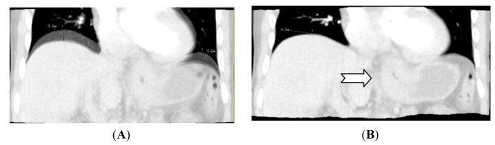
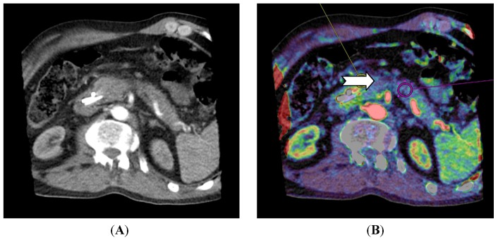
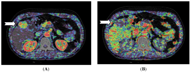
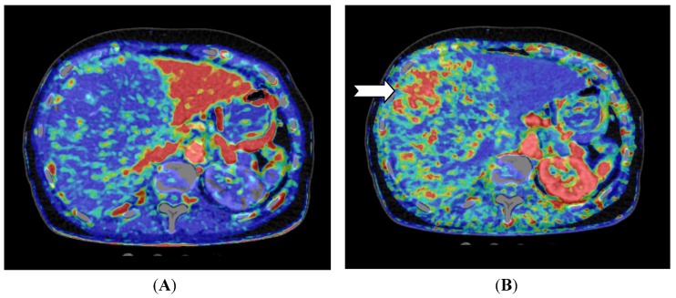
Computed Tomography (CT) Perfusion is an evolving method to visualize perfusion in organs and tissue. With the introduction of multidetector CT scanners, it is now possible to cover up to 16 cm in one rotation, and thereby making it possible to scan entire organs such as the liver with a fixed table position. Advances in reconstruction algorithms make it possible to reduce the radiation dose for each examination to acceptable levels. Regarding abdominal imaging, CT perfusion is still considered a research tool, but several studies have proven it as a reliable non-invasive technique for assessment of vascularity. CT perfusion has also been used for tumor characterization, staging of disease, response evaluation of newer drugs targeted towards angiogenesis and as a method for early detection of recurrence after radiation and embolization. There are several software solutions available on the market today based on different perfusion algorithms. However, there is no consensus on which protocol and algorithm to use for specific organs. In this article, the authors give an introduction to CT perfusion in abdominal imaging introducing technical aspects for calculation of perfusion parameters, and considerations on patient preparation. This article also contains clinical cases to illustrate the use of CT perfusion in abdominal imaging.

摘要

计算机断层扫描(CT)灌注是一种用于可视化器官和组织灌注的新兴方法。随着多排 CT 扫描仪的引入,现在可以在一次旋转中覆盖多达 16 厘米的范围,从而可以使用固定的台位扫描整个器官,如肝脏。重建算法的进步使得每个检查的辐射剂量都可以降低到可接受的水平。在腹部成像方面,CT 灌注仍然被认为是一种研究工具,但已有多项研究证明其是一种可靠的、非侵入性的血管评估技术。CT 灌注也已用于肿瘤特征描述、疾病分期、针对血管生成的新型药物的反应评估以及作为放射和栓塞后早期复发检测的方法。目前市场上有几种基于不同灌注算法的软件解决方案。然而,对于特定器官,使用哪种方案和算法还没有共识。本文的作者在腹部成像中介绍了 CT 灌注,介绍了计算灌注参数的技术方面,以及对患者准备的考虑。本文还包含了临床病例,以说明 CT 灌注在腹部成像中的应用。

https://cdn.ncbi.nlm.nih.gov/pmc/blobs/8e8c/4665537/495b2b809b19/diagnostics-03-00261-g004.jpg
https://cdn.ncbi.nlm.nih.gov/pmc/blobs/8e8c/4665537/eebbb23db9cd/diagnostics-03-00261-g001.jpg
https://cdn.ncbi.nlm.nih.gov/pmc/blobs/8e8c/4665537/a564ca008165/diagnostics-03-00261-g002.jpg
https://cdn.ncbi.nlm.nih.gov/pmc/blobs/8e8c/4665537/aa37159b3bf3/diagnostics-03-00261-g003.jpg
https://cdn.ncbi.nlm.nih.gov/pmc/blobs/8e8c/4665537/495b2b809b19/diagnostics-03-00261-g004.jpg

相似文献

1
Computed Tomography (CT) Perfusion in Abdominal Cancer: Technical Aspects.腹部癌症的计算机断层扫描 (CT) 灌注:技术方面。
Diagnostics (Basel). 2013 Apr 3;3(2):261-70. doi: 10.3390/diagnostics3020261.
2
More advantages in detecting bone and soft tissue metastases from prostate cancer using F-PSMA PET/CT.使用F-PSMA PET/CT检测前列腺癌骨和软组织转移方面有更多优势。
Hell J Nucl Med. 2019 Jan-Apr;22(1):6-9. doi: 10.1967/s002449910952. Epub 2019 Mar 7.
3
CT perfusion of the liver: principles and applications in oncology.肝脏 CT 灌注:肿瘤学中的原理与应用。
Radiology. 2014 Aug;272(2):322-44. doi: 10.1148/radiol.14130091.
4
Qualitative and quantitative evaluation of rigid and deformable motion correction algorithms using dual-energy CT images in view of application to CT perfusion measurements in abdominal organs affected by breathing motion.鉴于在受呼吸运动影响的腹部器官的CT灌注测量中的应用,使用双能CT图像对刚性和可变形运动校正算法进行定性和定量评估。
Br J Radiol. 2015 Feb;88(1046):20140683. doi: 10.1259/bjr.20140683. Epub 2014 Dec 3.
5
[Research Progress of Low Radiation Dose Computed Tomography Perfusion of the Liver].[肝脏低辐射剂量计算机断层扫描灌注的研究进展]
Sheng Wu Yi Xue Gong Cheng Xue Za Zhi. 2016 Apr;33(2):400-4.
6
Quantitative computed tomography liver perfusion imaging using dynamic spiral scanning with variable pitch: feasibility and initial results in patients with cancer metastases.应用动态螺旋扫描可变螺距技术的定量 CT 肝脏灌注成像:在转移性癌症患者中的可行性及初步结果。
Invest Radiol. 2010 Jul;45(7):419-26. doi: 10.1097/RLI.0b013e3181e1937b.
7
CT perfusion of renal cell carcinoma: impact of volume coverage on quantitative analysis.CT 灌注成像在肾细胞癌中的应用:容积覆盖范围对定量分析的影响。
Invest Radiol. 2012 Jan;47(1):33-40. doi: 10.1097/RLI.0b013e31822598c3.
8
Colorectal cancer. Radiologic staging.结直肠癌。放射学分期。
Radiol Clin North Am. 1997 Mar;35(2):457-85.
9
Multidetector CT of blunt abdominal trauma.多排 CT 平扫在钝性腹部创伤中的应用。
Radiology. 2012 Dec;265(3):678-93. doi: 10.1148/radiol.12120354.
10
Differences in perfusion CT parameter values with commercial software upgrades: a preliminary report about algorithm consistency and stability.商用软件升级后灌注CT参数值的差异:关于算法一致性和稳定性的初步报告
Acta Radiol. 2013 Sep;54(7):805-11. doi: 10.1177/0284185113484643. Epub 2013 Apr 30.

引用本文的文献

1
High ischemic deficit in computed tomography perfusion is a risk factor for cerebral vascular ischemic events in Takayasu arteritis: a prospective observational study.计算机断层扫描灌注中高缺血性缺损是大动脉炎脑血管缺血事件的危险因素:一项前瞻性观察研究。
BMC Rheumatol. 2025 Aug 13;9(1):100. doi: 10.1186/s41927-025-00551-6.
2
A Systematic Review on the Role of the Perfusion Computed Tomography in Abdominal Cancer.灌注计算机断层扫描在腹部癌症中作用的系统评价
Dose Response. 2021 Nov 24;19(4):15593258211056199. doi: 10.1177/15593258211056199. eCollection 2021 Oct-Dec.
3
Advancement in Diagnostic Imaging of Thymic Tumors.

本文引用的文献

1
Comparison of free breathing versus breath-hold in perfusion imaging using dynamic volume CT.使用动态容积 CT 进行灌注成像时,自由呼吸与屏气的比较。
Insights Imaging. 2012 Aug;3(4):323-8. doi: 10.1007/s13244-012-0169-9. Epub 2012 May 1.
2
Hepatic CT perfusion measurements: a feasibility study for radiation dose reduction using new image reconstruction method.肝脏 CT 灌注测量:使用新的图像重建方法降低辐射剂量的可行性研究。
Eur J Radiol. 2012 Nov;81(11):3048-54. doi: 10.1016/j.ejrad.2012.04.024. Epub 2012 May 19.
3
Perfusion CT in colorectal cancer: comparison of perfusion parameters with tumor grade and microvessel density.
胸腺肿瘤诊断成像的进展
Cancers (Basel). 2021 Jul 18;13(14):3599. doi: 10.3390/cancers13143599.
4
Can Computed Tomography Perfusion Predict Treatment Response After Prostate Artery Embolization: A Feasibility Study.计算机断层扫描灌注能否预测前列腺动脉栓塞术后的治疗反应:一项可行性研究。
Diagnostics (Basel). 2020 May 15;10(5):304. doi: 10.3390/diagnostics10050304.
5
Evaluation of efficacy of transcatheter arterial chemoembolization combined with computed tomography-guided radiofrequency ablation for hepatocellular carcinoma using magnetic resonance diffusion weighted imaging and computed tomography perfusion imaging: A prospective study.利用磁共振扩散加权成像和计算机断层扫描灌注成像评估经导管动脉化疗栓塞联合计算机断层扫描引导下射频消融治疗肝细胞癌的疗效:一项前瞻性研究。
Medicine (Baltimore). 2017 Jan;96(3):e5518. doi: 10.1097/MD.0000000000005518.
6
Dynamic Contrast-Enhanced CT in Patients with Pancreatic Cancer.动态对比增强 CT 在胰腺癌患者中的应用。
Diagnostics (Basel). 2016 Sep 6;6(3):34. doi: 10.3390/diagnostics6030034.
7
Assessing Tumor Response to Treatment in Patients with Lung Cancer Using Dynamic Contrast-Enhanced CT.使用动态对比增强 CT 评估肺癌患者的肿瘤治疗反应。
Diagnostics (Basel). 2016 Jul 21;6(3):28. doi: 10.3390/diagnostics6030028.
8
A Bayesian Nonparametric Approach for Functional Data Classification with Application to Hepatic Tissue Characterization.一种用于功能数据分类的贝叶斯非参数方法及其在肝组织特征描述中的应用
Cancer Inform. 2016 May 29;14(Suppl 5):151-62. doi: 10.4137/CIN.S31933. eCollection 2015.
9
Interobserver and Intraobserver Reproducibility with Volume Dynamic Contrast Enhanced Computed Tomography (DCE-CT) in Gastroesophageal Junction Cancer.胃食管结合部癌的容积动态对比增强 CT(DCE-CT)的观察者间和观察者内可重复性。
Diagnostics (Basel). 2016 Feb 1;6(1):8. doi: 10.3390/diagnostics6010008.
10
Computed tomography (CT) perfusion as an early predictive marker for treatment response to neoadjuvant chemotherapy in gastroesophageal junction cancer and gastric cancer--a prospective study.计算机断层扫描(CT)灌注作为胃食管交界癌和胃癌新辅助化疗治疗反应的早期预测标志物——一项前瞻性研究。
PLoS One. 2014 May 20;9(5):e97605. doi: 10.1371/journal.pone.0097605. eCollection 2014.
结直肠癌灌注 CT:灌注参数与肿瘤分级和微血管密度的比较。
Korean J Radiol. 2012 Jan-Feb;13 Suppl 1(Suppl 1):S89-97. doi: 10.3348/kjr.2012.13.S1.S89. Epub 2012 Apr 23.
4
Pancreatic Perfusion CT in Early Stage of Severe Acute Pancreatitis.重症急性胰腺炎早期的胰腺灌注CT
Int J Inflam. 2012;2012:497386. doi: 10.1155/2012/497386. Epub 2012 Feb 15.
5
Brain perfusion CT compared with15O-H2O-PET in healthy subjects.脑灌注 CT 与 15O-H2O-PET 在健康受试者中的比较。
EJNMMI Res. 2011 Nov 18;1(1):28. doi: 10.1186/2191-219X-1-28.
6
CT image quality improvement using Adaptive Iterative Dose Reduction with wide-volume acquisition on 320-detector CT.利用 320 排探测器 CT 宽体采集自适应迭代剂量降低技术改善 CT 图像质量。
Eur Radiol. 2012 Feb;22(2):295-301. doi: 10.1007/s00330-011-2271-7. Epub 2011 Sep 17.
7
Deconvolution-Based CT and MR Brain Perfusion Measurement: Theoretical Model Revisited and Practical Implementation Details.基于去卷积的CT和MR脑灌注测量:理论模型再探讨及实际实施细节
Int J Biomed Imaging. 2011;2011:467563. doi: 10.1155/2011/467563. Epub 2011 Aug 28.
8
CT hepatic perfusion measurement: comparison of three analytic methods.CT 肝灌注测量:三种分析方法的比较。
Eur J Radiol. 2012 Sep;81(9):2075-9. doi: 10.1016/j.ejrad.2011.07.003. Epub 2011 Jul 29.
9
Reproducibility of CT perfusion parameters in liver tumors and normal liver.肝脏肿瘤和正常肝脏 CT 灌注参数的可重复性。
Radiology. 2011 Sep;260(3):762-70. doi: 10.1148/radiol.11110331. Epub 2011 Jul 25.
10
Assessment of tumor vascularization in pancreatic adenocarcinoma using 128-slice perfusion computed tomography imaging.使用128层灌注计算机断层扫描成像评估胰腺腺癌中的肿瘤血管生成情况。
J Comput Assist Tomogr. 2011 Jul-Aug;35(4):434-8. doi: 10.1097/RCT.0b013e318223f0c5.