• 文献检索
  • 文档翻译
  • 深度研究
  • 学术资讯
  • Suppr Zotero 插件Zotero 插件
  • 邀请有礼
  • 套餐&价格
  • 历史记录
应用&插件
Suppr Zotero 插件Zotero 插件浏览器插件Mac 客户端Windows 客户端微信小程序
定价
高级版会员购买积分包购买API积分包
服务
文献检索文档翻译深度研究API 文档MCP 服务
关于我们
关于 Suppr公司介绍联系我们用户协议隐私条款
关注我们

Suppr 超能文献

核心技术专利:CN118964589B侵权必究
粤ICP备2023148730 号-1Suppr @ 2026

文献检索

告别复杂PubMed语法,用中文像聊天一样搜索,搜遍4000万医学文献。AI智能推荐,让科研检索更轻松。

立即免费搜索

文件翻译

保留排版,准确专业,支持PDF/Word/PPT等文件格式,支持 12+语言互译。

免费翻译文档

深度研究

AI帮你快速写综述,25分钟生成高质量综述,智能提取关键信息,辅助科研写作。

立即免费体验

感音神经性听力损失的影像学:内耳和桥小脑角疾病的基于模式的方法。

Imaging of sensorineural hearing loss: a pattern-based approach to diseases of the inner ear and cerebellopontine angle.

机构信息

Department of Radiology, Leiden University Medical Center, PO Box 9600, 2300 RC, Leiden, The Netherlands,

出版信息

Insights Imaging. 2012 Apr;3(2):139-53. doi: 10.1007/s13244-011-0134-z. Epub 2011 Dec 9.

DOI:10.1007/s13244-011-0134-z
PMID:22696040
原文链接:https://pmc.ncbi.nlm.nih.gov/articles/PMC3314739/
Abstract

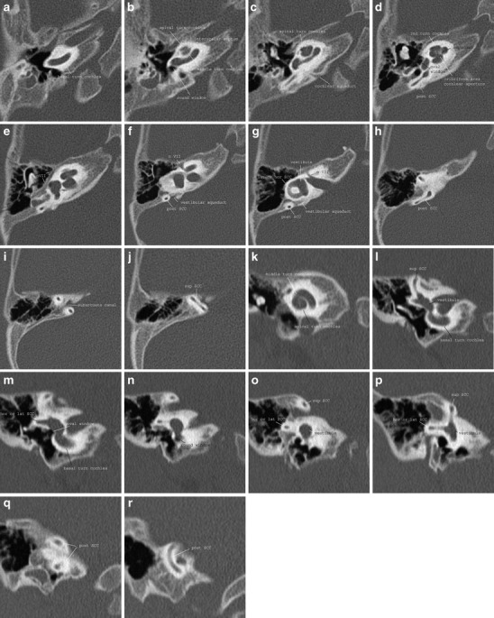
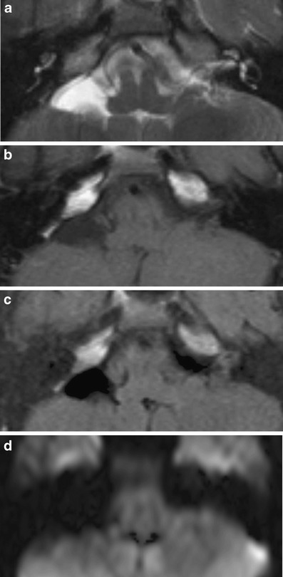
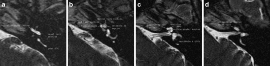
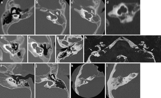
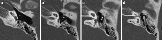
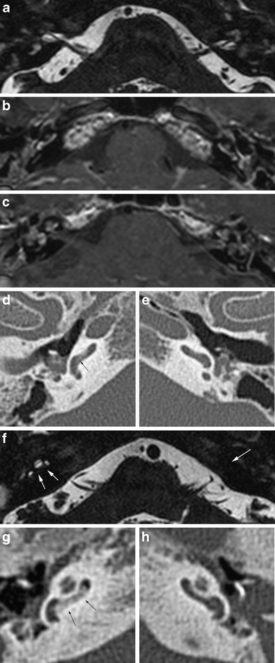
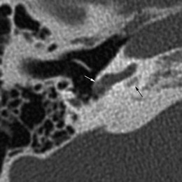
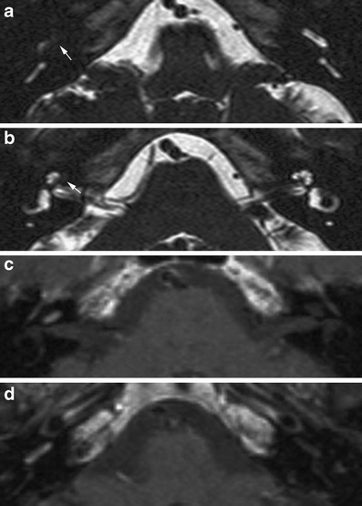
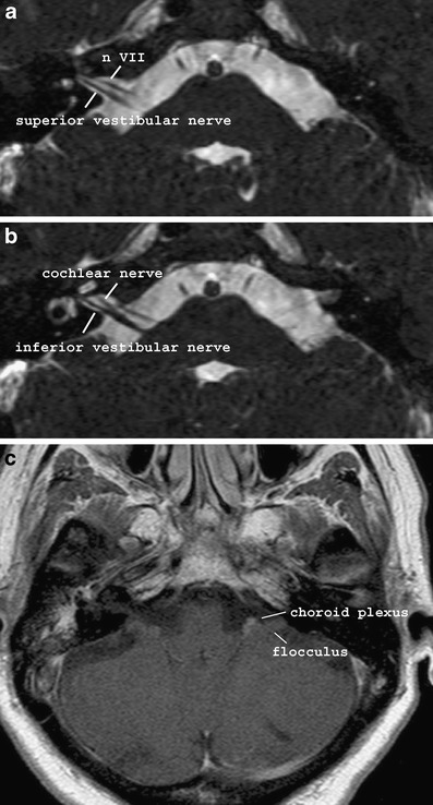
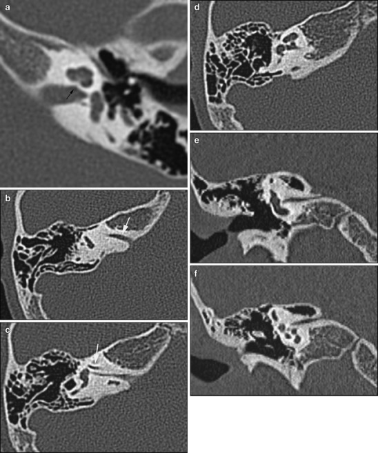
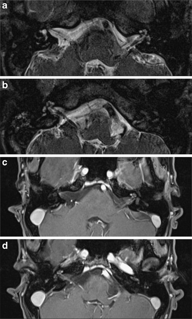
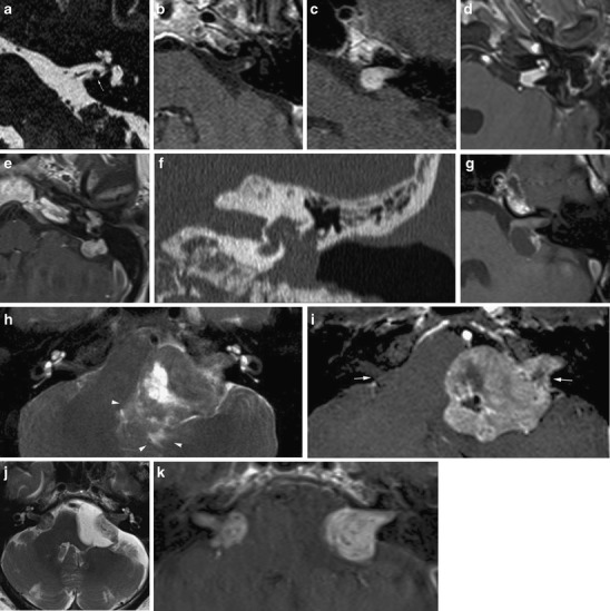
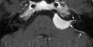
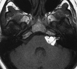
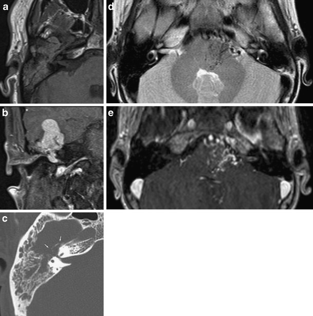
An overview is presented of the common and uncommon diseases of the inner ear and of the cochleovestibular nerve within the internal acoustic meatus and cerebellopontine angle cistern causing sensorineural deafness.An imaging-pattern-based approach is used to help detect disease and narrow the differential diagnosis. Main Messages • The most common soft tissue mass lesions in the cerebellopontine angle are schwannoma and meningioma. • Contrast-enhanced MRI may reveal clinically unsuspected inflammatory, auto-immune or tumoural disease. • Hearing loss may be caused by infection, inflammation or, rarely, perineural tumour spread along the cochleovestibular nerve. • Labyrinthitis may lead to rapidly progressive ossification of the labyrinth.

摘要

本文对内耳和内听道及桥小脑角池内的耳蜗前庭神经的常见和罕见疾病进行了概述,这些疾病可导致感音神经性聋。采用基于影像学表现的方法有助于发现疾病并缩小鉴别诊断范围。

主要信息

• 桥小脑角最常见的软组织肿块病变是神经鞘瘤和脑膜瘤。

• 增强 MRI 可能会显示出临床上未怀疑的炎症、自身免疫或肿瘤性疾病。

• 听力损失可由感染、炎症引起,或很少由沿耳蜗前庭神经的神经周肿瘤扩散引起。

• 迷路炎可导致迷路迅速进行性骨化。

https://cdn.ncbi.nlm.nih.gov/pmc/blobs/7091/3314739/7ec3efaf70b6/13244_2011_134_Fig17_HTML.jpg
https://cdn.ncbi.nlm.nih.gov/pmc/blobs/7091/3314739/a9de5ad756a4/13244_2011_134_Fig1_HTML.jpg
https://cdn.ncbi.nlm.nih.gov/pmc/blobs/7091/3314739/1bf57f49ebaf/13244_2011_134_Fig2_HTML.jpg
https://cdn.ncbi.nlm.nih.gov/pmc/blobs/7091/3314739/91cf6458644d/13244_2011_134_Fig3_HTML.jpg
https://cdn.ncbi.nlm.nih.gov/pmc/blobs/7091/3314739/8a7f4fed4ed8/13244_2011_134_Fig4_HTML.jpg
https://cdn.ncbi.nlm.nih.gov/pmc/blobs/7091/3314739/003f64a697d6/13244_2011_134_Fig5_HTML.jpg
https://cdn.ncbi.nlm.nih.gov/pmc/blobs/7091/3314739/47f362c8cf86/13244_2011_134_Fig6_HTML.jpg
https://cdn.ncbi.nlm.nih.gov/pmc/blobs/7091/3314739/0fab34210150/13244_2011_134_Fig7_HTML.jpg
https://cdn.ncbi.nlm.nih.gov/pmc/blobs/7091/3314739/b0d5a163d103/13244_2011_134_Fig8_HTML.jpg
https://cdn.ncbi.nlm.nih.gov/pmc/blobs/7091/3314739/21b7913e9008/13244_2011_134_Fig9_HTML.jpg
https://cdn.ncbi.nlm.nih.gov/pmc/blobs/7091/3314739/01270a643f27/13244_2011_134_Fig10_HTML.jpg
https://cdn.ncbi.nlm.nih.gov/pmc/blobs/7091/3314739/73c9b05e1267/13244_2011_134_Fig11_HTML.jpg
https://cdn.ncbi.nlm.nih.gov/pmc/blobs/7091/3314739/428f63d51864/13244_2011_134_Fig12_HTML.jpg
https://cdn.ncbi.nlm.nih.gov/pmc/blobs/7091/3314739/eee8eeab6384/13244_2011_134_Fig13_HTML.jpg
https://cdn.ncbi.nlm.nih.gov/pmc/blobs/7091/3314739/277c0333c220/13244_2011_134_Fig14_HTML.jpg
https://cdn.ncbi.nlm.nih.gov/pmc/blobs/7091/3314739/cc13a4b86aec/13244_2011_134_Fig15_HTML.jpg
https://cdn.ncbi.nlm.nih.gov/pmc/blobs/7091/3314739/384a82fe9980/13244_2011_134_Fig16_HTML.jpg
https://cdn.ncbi.nlm.nih.gov/pmc/blobs/7091/3314739/7ec3efaf70b6/13244_2011_134_Fig17_HTML.jpg

相似文献

1
Imaging of sensorineural hearing loss: a pattern-based approach to diseases of the inner ear and cerebellopontine angle.感音神经性听力损失的影像学:内耳和桥小脑角疾病的基于模式的方法。
Insights Imaging. 2012 Apr;3(2):139-53. doi: 10.1007/s13244-011-0134-z. Epub 2011 Dec 9.
2
Flocculus Herniation and Hearing Disturbance Induced by Remote Tentorial Meningioma: A Case Report.远隔部位小脑幕脑膜瘤所致小脑绒球疝及听力障碍:1例报告
NMC Case Rep J. 2018 Sep 20;5(4):73-76. doi: 10.2176/nmccrj.cr.2018-0059. eCollection 2018 Oct.
3
Sensorineural hearing loss: more than meets the eye?感音神经性听力损失:表象之下另有隐情?
AJNR Am J Neuroradiol. 1993 Jan-Feb;14(1):37-45.
4
[Computed tomography and magnetic resonance imaging of acquired abnormalities of the inner ear and cerebello-pontine angle].[内耳及桥小脑角后天性异常的计算机断层扫描与磁共振成像]
Radiologe. 2003 Mar;43(3):213-8. doi: 10.1007/s00117-003-0872-0.
5
[MRI in progressive sensorineural hearing loss].[磁共振成像在进行性感音神经性听力损失中的应用]
Acta Otorhinolaryngol Ital. 1998 Aug;18(4 Suppl 59):80-6.
6
An arteriovenous malformation within the internal acoustic meatus and cerebellopontine angle cistern.内耳道及桥小脑角池内的动静脉畸形。
J Laryngol Otol. 2011 Dec;125(12):1275-8. doi: 10.1017/S0022215111002155. Epub 2011 Aug 16.
7
Magnetic resonance examination of the inner ear and cerebellopontine angle in patients with vertigo and/or abnormal findings at vestibular testing.对眩晕患者和/或前庭测试有异常发现的患者进行内耳及桥小脑角的磁共振检查。
Acta Otolaryngol Suppl. 1994;513:15-27. doi: 10.3109/00016489409127322.
8
Detection of acoustic schwannoma: use of constructive interference in the steady state three-dimensional MR.听神经鞘瘤的检测:稳态三维磁共振成像中相长干涉的应用
AJNR Am J Neuroradiol. 1996 Aug;17(7):1219-25.
9
Cerebellopontine Angle Anaplastic Ganglioglioma Masquerading as Vestibular Schwannoma: Unusual Entity.伪装成前庭神经鞘瘤的桥小脑角间变性节细胞胶质瘤:罕见病例
World Neurosurg. 2018 Sep;117:221-224. doi: 10.1016/j.wneu.2018.06.054. Epub 2018 Jun 19.
10
Nationwide clinico-epidemiological treatment analysis of adult patients with tumors of cerebellopontine angle and internal acoustic meatus in Poland during 2011-2020.波兰 2011-2020 年成人桥小脑角和内听道肿瘤患者的全国临床流行病学治疗分析。
BMC Public Health. 2023 Sep 6;23(1):1735. doi: 10.1186/s12889-023-16551-5.

引用本文的文献

1
The State of High-Resolution Imaging of the Human Inner Ear: A Look Into the Black Box.人类内耳的高分辨率成像现状:窥探黑匣子
Adv Sci (Weinh). 2025 Jul;12(28):e00556. doi: 10.1002/advs.202500556. Epub 2025 Jun 5.
2
Imaging in sensorineural and conductive hearing loss-an educational review.感音神经性和传导性听力损失的影像学——一篇教育性综述
Radiol Med. 2025 Jan;130(1):46-62. doi: 10.1007/s11547-024-01922-8. Epub 2024 Nov 12.
3
Guidelines for magnetic resonance imaging in pediatric head and neck pathologies: a multicentre international consensus paper.

本文引用的文献

1
Aberrant course of the intracranial facial nerve in cases of atresia of the internal auditory canal (IAC).内听道闭锁时颅内面神经的异常走行。
Neuroradiology. 2011 Sep;53(9):681-7. doi: 10.1007/s00234-011-0862-2. Epub 2011 Mar 30.
2
A hemorrhagic vestibular schwannoma presenting with rapid neurologic decline: a case report.表现为快速神经功能下降的出血性前庭神经鞘瘤:病例报告。
Laryngoscope. 2010;120 Suppl 4:S204. doi: 10.1002/lary.21671.
3
Dutch Cochlear Implant Group (CI-ON) consensus protocol on postmeningitis hearing evaluation and treatment.
儿童头颈部病变磁共振成像指南:多中心国际共识文件。
Neuroradiology. 2022 Jun;64(6):1081-1100. doi: 10.1007/s00234-022-02950-9. Epub 2022 Apr 23.
4
Unilateral sudden sensorineural hearing loss with vertigo as a first symptom of bilateral internal auditory canal metastases: a case report.单侧突发性感觉神经性耳聋伴眩晕作为双侧内听道转移的首发症状:病例报告。
J Int Med Res. 2021 Oct;49(10):3000605211053553. doi: 10.1177/03000605211053553.
5
The role of diffusion tensor imaging in idiopathic sensorineural hearing loss: is it significant?扩散张量成像在特发性感音神经性听力损失中的作用:它重要吗?
Pol J Radiol. 2021 Aug 9;86:e474-e480. doi: 10.5114/pjr.2021.108372. eCollection 2021.
6
Flocculus Herniation and Hearing Disturbance Induced by Remote Tentorial Meningioma: A Case Report.远隔部位小脑幕脑膜瘤所致小脑绒球疝及听力障碍:1例报告
NMC Case Rep J. 2018 Sep 20;5(4):73-76. doi: 10.2176/nmccrj.cr.2018-0059. eCollection 2018 Oct.
7
Hearing loss in patients with scleroderma: associations with clinical manifestations and capillaroscopy.硬皮病患者的听力损失:与临床表现和毛细血管镜检查的关联。
Clin Rheumatol. 2018 Sep;37(9):2439-2446. doi: 10.1007/s10067-018-4162-7. Epub 2018 Jun 2.
8
Atypical bilateral cerebellopontine angle medulloblastoma: differential diagnosis, immunohistochemical features and radiological presentation.非典型双侧小脑桥脑角髓母细胞瘤:鉴别诊断、免疫组化特征及影像学表现
Contemp Oncol (Pozn). 2017;21(3):249-253. doi: 10.5114/wo.2017.69593. Epub 2017 Sep 29.
9
Visualization of human inner ear anatomy with high-resolution MR imaging at 7T: initial clinical assessment.7T高分辨率磁共振成像对人内耳解剖结构的可视化:初步临床评估
AJNR Am J Neuroradiol. 2015 Feb;36(2):378-83. doi: 10.3174/ajnr.A4084. Epub 2014 Aug 21.
荷兰耳蜗植入物专家组(CI-ON)关于脑膜炎后听力评估和治疗的共识方案。
Otol Neurotol. 2010 Oct;31(8):1281-6. doi: 10.1097/MAO.0b013e3181f1fc58.
4
Spontaneous subarachnoid haemorrhage due to acoustic neurinoma. Case report and review of the literature.听神经瘤导致的自发性蛛网膜下腔出血。病例报告及文献复习
Br J Neurosurg. 2010 Feb;24(1):82-3. doi: 10.3109/02688690903506085.
5
Vestibular Schwannomas presenting with haemorrhage: clinical presentation and histopathological evaluation of an unusual entity.
Br J Neurosurg. 2009 Aug;23(4):431-6. doi: 10.1080/02688690902968828.
6
Genetics of aminoglycoside-induced and prelingual non-syndromic mitochondrial hearing impairment: a review.氨基糖苷类药物所致及语前非综合征性线粒体听力障碍的遗传学:综述
Int J Audiol. 2008 Nov;47(11):702-7. doi: 10.1080/14992020802215862.
7
Imaging of intralabyrinthine schwannomas: a retrospective study of 52 cases with emphasis on lesion growth.迷路内神经鞘瘤的影像学表现:一项对52例病例的回顾性研究,重点关注病变生长情况。
AJNR Am J Neuroradiol. 2008 May;29(5):898-905. doi: 10.3174/ajnr.A1026. Epub 2008 Mar 5.
8
Intratumoral microhemorrhages on T2*-weighted gradient-echo imaging helps differentiate vestibular schwannoma from meningioma.T2*加权梯度回波成像上的瘤内微出血有助于鉴别前庭神经鞘瘤和脑膜瘤。
AJNR Am J Neuroradiol. 2008 Mar;29(3):552-7. doi: 10.3174/ajnr.A0887. Epub 2007 Dec 13.
9
Deafness after bacterial meningitis: an emergency for early imaging and cochlear implant surgery.
Otolaryngol Head Neck Surg. 2005 Dec;133(6):995-6. doi: 10.1016/j.otohns.2005.03.036.
10
Inner ear extension of vestibular schwannomas.前庭神经鞘瘤的内耳延伸
Laryngoscope. 2003 Sep;113(9):1605-8. doi: 10.1097/00005537-200309000-00037.