Hegde Harihar V, Bhat Ravi L, Shanbag Raghunath D, Bharat Mp, Rao P Raghavendra
Department of Anaesthesiology, SDM College of Medical Sciences and Hospital, Dharwad, Karnataka, India.
Indian J Anaesth. 2012 Mar;56(2):171-4. doi: 10.4103/0019-5049.96338.
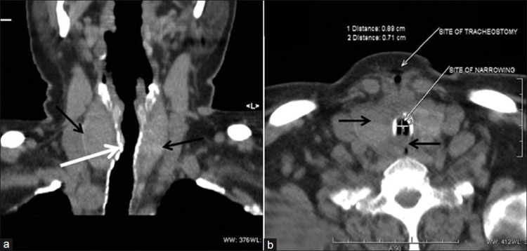
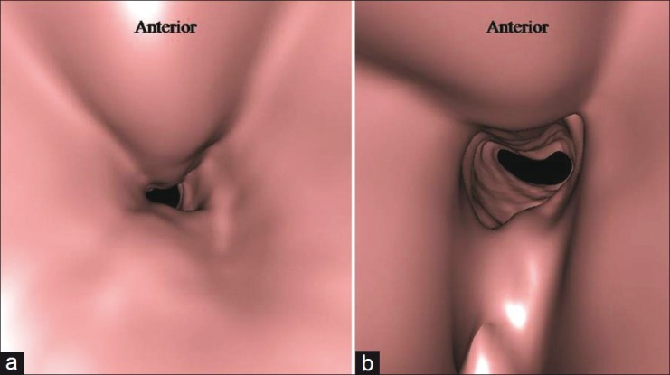
Patients with chronic obstructive pulmonary disease (COPD) are susceptible to airway malacia, which may be unmasked following mechanical ventilation or tracheostomy decannulation. Dynamic imaging of central airways, a non-invasive test as effective as bronchoscopy to diagnose airway malacia, has increased the recognition of this disorder. We describe a 70-year-old woman admitted with adult respiratory distress syndrome. She had cardiorespiratory arrest on admission, from which she was successfully resuscitated. She had obesity, hypertension, diabetes mellitus, recurrent ventricular tachycardia, sarcoidosis with interstitial lung disease and COPD. She received short-term (18 days) mechanical ventilation with tracheostomy and developed respiratory distress following tracheostomy decannulation.
慢性阻塞性肺疾病(COPD)患者易患气道软化症,在机械通气或气管切开拔管后可能会显现出来。中央气道的动态成像作为一种与支气管镜检查诊断气道软化症同样有效的非侵入性检查,提高了对这种疾病的认识。我们描述了一名70岁因成人呼吸窘迫综合征入院的女性。她入院时发生心肺骤停,后成功复苏。她患有肥胖症、高血压、糖尿病、复发性室性心动过速、结节病合并间质性肺病和COPD。她接受了短期(18天)带气管切开的机械通气,并在气管切开拔管后出现呼吸窘迫。