Neuroscience Research Centre, Shohda Hospital, Tabriz University of Medical Sciences, Tabriz, Iran.
Eur Spine J. 2013 May;22 Suppl 3(Suppl 3):S329-36. doi: 10.1007/s00586-012-2373-1. Epub 2012 Jun 16.
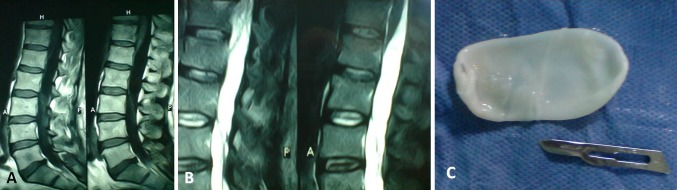
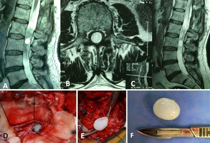
Primary intradural extramedullary hydatid cyst is a rare form of parasitic infection, causing focal neurological signs, commonly observed in sheep-raising areas of the world. We report a rare case of intradural, extramedullary spinal cyst, which we had misdiagnosis in the first surgery, because of rarity of the case. A 55-year-old man presented to our hospital in August 2008. He was admitted to our clinic because of lumbar pain of increasing severity and progressive difficulty with walking and stiffness of both lower limbs, which had lasted for 1 month. On the basis of imaging results, arachnoid cyst of the lumbar spine was diagnosed. Due to rapid progression of the patient's symptoms toward spastic paraplegia, he underwent an emergency surgical decompression procedure. The patient underwent exploratory surgery using a posterior approach. A L1-L2 laminectomy was performed. After opening the dura, an intradural extramedullary cystic mass was determined. The surgical specimen measured 6 × 2 cm and was described as a whitish, pearl-like, semitranslucent, cystic material, which was thought to be parasitic. Surgery has to be followed by albendazole therapy.
原发性硬脊膜外髓内包虫囊肿是一种罕见的寄生虫感染形式,引起局灶性神经体征,常见于世界上的绵羊养殖区。我们报告一例罕见的硬脊膜外脊髓囊肿,由于该病例罕见,我们在第一次手术中误诊。一名 55 岁男性于 2008 年 8 月就诊于我院。他因腰痛逐渐加重、进行性行走困难和双下肢僵硬而入住我院,症状持续了 1 个月。根据影像学结果,诊断为腰骶部蛛网膜囊肿。由于患者的症状迅速进展为痉挛性截瘫,他接受了紧急手术减压。患者采用后路进行探查性手术。进行了 L1-L2 椎板切除术。打开硬脑膜后,确定为硬脊膜外髓内囊性肿块。手术标本大小为 6×2cm,呈白色、珍珠样、半透明的囊性物质,被认为是寄生虫。手术治疗后必须进行阿苯达唑治疗。