The Centre of Inflammation and Metabolism at Department of Infectious Diseases and Copenhagen Muscle Research Centre, Rigshospitalet, The Faculty of Health Sciences, University of Copenhagen, Copenhagen, Denmark.
PLoS One. 2012;7(6):e39657. doi: 10.1371/journal.pone.0039657. Epub 2012 Jun 26.
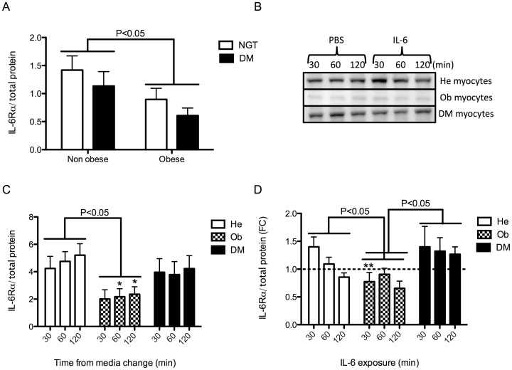
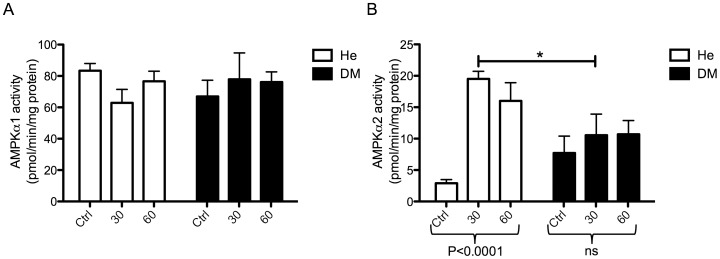
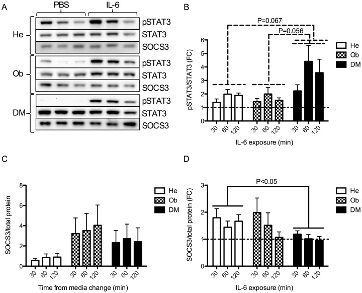
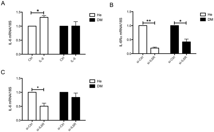
Obesity and type 2 diabetes are associated with chronically elevated systemic levels of IL-6, a pro-inflammatory cytokine with a role in skeletal muscle metabolism that signals through the IL-6 receptor (IL-6Rα). We hypothesized that skeletal muscle in obesity-associated type 2 diabetes develops a resistance to IL-6. By utilizing western blot analysis, we demonstrate that IL-6Rα protein was down regulated in skeletal muscle biopsies from obese persons with and without type 2 diabetes. To further investigate the status of IL-6 signaling in skeletal muscle in obesity-associated type 2 diabetes, we isolated satellite cells from skeletal muscle of people that were healthy (He), obese (Ob) or were obese and had type 2 diabetes (DM), and differentiated them in vitro into myocytes. Down-regulation of IL-6Rα was conserved in Ob myocytes. In addition, acute IL-6 administration for 30, 60 and 120 minutes, resulted in a down-regulation of IL-6Rα protein in Ob myocytes compared to both He myocytes (P<0.05) and DM myocytes (P<0.05). Interestingly, there was a strong time-dependent regulation of IL-6Rα protein in response to IL-6 (P<0.001) in He myocytes, not present in the other groups. Assessing downstream signaling, DM, but not Ob myocytes demonstrated a trend towards an increased protein phosphorylation of STAT3 in DM myocytes (P = 0.067) accompanied by a reduced SOCS3 protein induction (P<0.05), in response to IL-6 administration. Despite this loss of negative control, IL-6 failed to increase AMPKα2 activity and IL-6 mRNA expression in DM myocytes. There was no difference in fusion capacity of myocytes between cell groups. Our data suggest that negative control of IL-6 signaling is increased in myocytes in obesity, whereas a dysfunctional IL-6 signaling is established further downstream of IL-6Rα in DM myocytes, possibly representing a novel mechanism by which skeletal muscle function is compromised in type 2 diabetes.
肥胖和 2 型糖尿病与系统中 IL-6 水平的慢性升高有关,IL-6 是一种促炎细胞因子,在骨骼肌代谢中起作用,并通过 IL-6 受体(IL-6Rα)发出信号。我们假设肥胖相关 2 型糖尿病患者的骨骼肌对 IL-6 产生了抗性。通过利用 Western blot 分析,我们证明了肥胖伴或不伴 2 型糖尿病患者的骨骼肌活检中 IL-6Rα 蛋白下调。为了进一步研究肥胖相关 2 型糖尿病患者骨骼肌中 IL-6 信号的状态,我们从健康(He)、肥胖(Ob)或肥胖且患有 2 型糖尿病(DM)的人的骨骼肌中分离卫星细胞,并在体外将其分化为肌细胞。Ob 肌细胞中 IL-6Rα 的下调是保守的。此外,急性给予 IL-630、60 和 120 分钟后,Ob 肌细胞中 IL-6Rα 蛋白的下调与 He 肌细胞(P<0.05)和 DM 肌细胞(P<0.05)相比。有趣的是,He 肌细胞中 IL-6Rα 蛋白对 IL-6 的时间依赖性调节较强,而其他组则没有。评估下游信号,DM 肌细胞而非 Ob 肌细胞表现出 IL-6 给药后 STAT3 蛋白磷酸化增加的趋势(P = 0.067),同时 SOCS3 蛋白诱导减少(P<0.05)。尽管这种负反馈控制丧失,但 IL-6 未能增加 DM 肌细胞中 AMPKα2 的活性和 IL-6 mRNA 的表达。细胞组之间肌细胞的融合能力没有差异。我们的数据表明,肥胖时肌细胞中 IL-6 信号的负反馈控制增加,而 DM 肌细胞中 IL-6Rα 下游的 IL-6 信号传递功能障碍,这可能代表了骨骼肌功能在 2 型糖尿病中受损的一种新机制。