Laboratory for Proteolytic Neuroscience, RIKEN Brain Science Institute, 2-1 Hirosawa, Wako-shi, Saitama 351-0198, Japan.
J Biol Chem. 2012 Aug 24;287(35):29362-72. doi: 10.1074/jbc.M112.340372. Epub 2012 Jul 5.
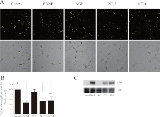
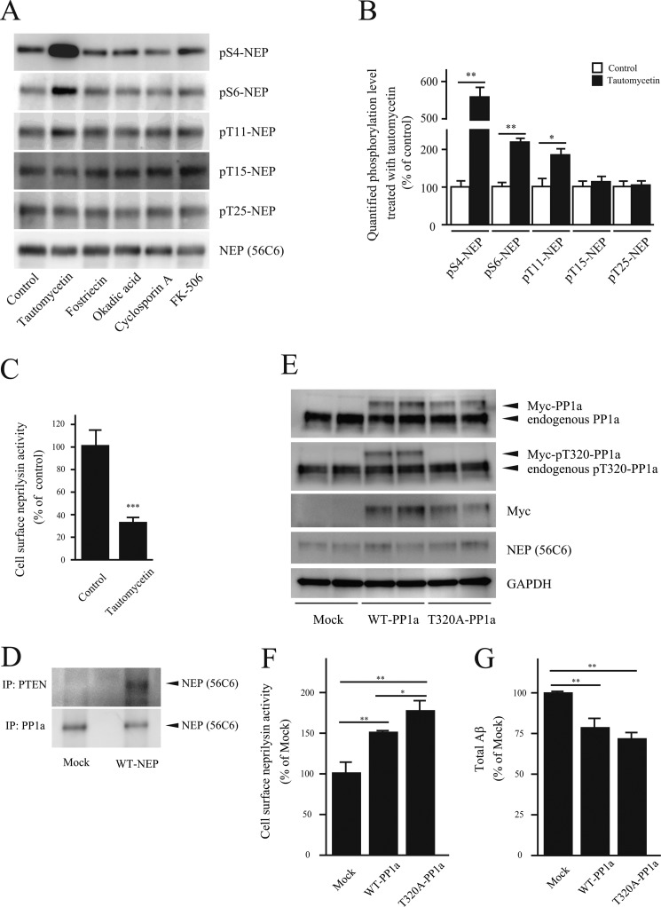
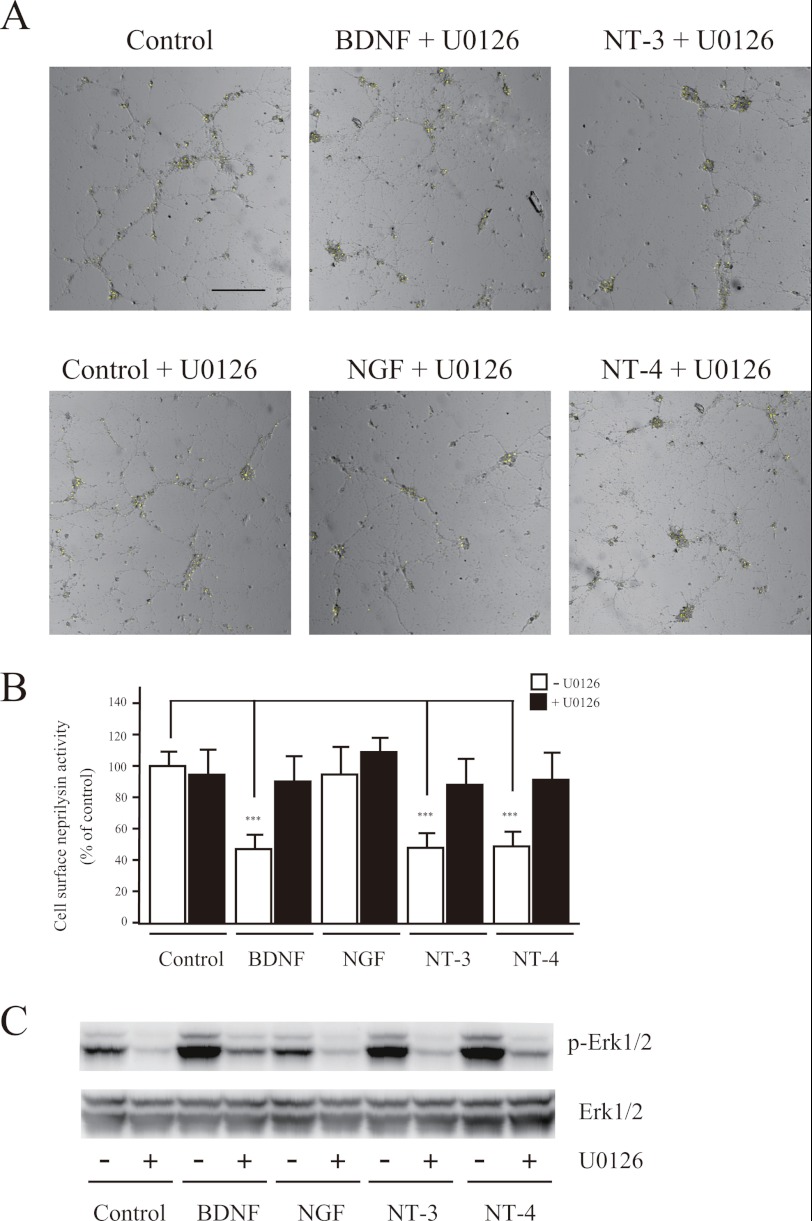
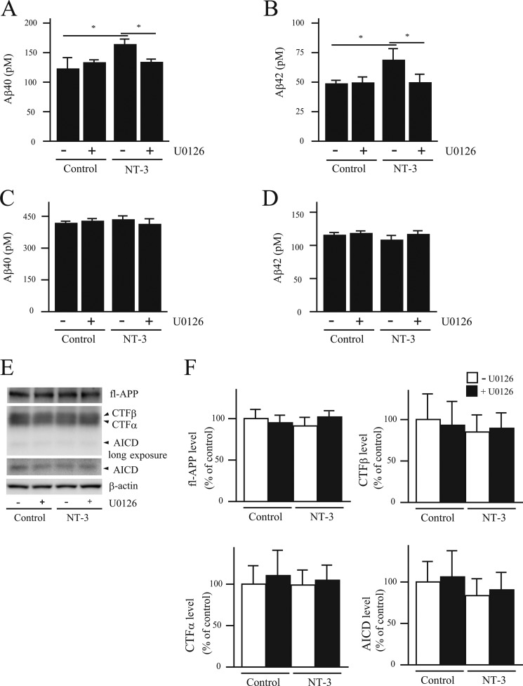
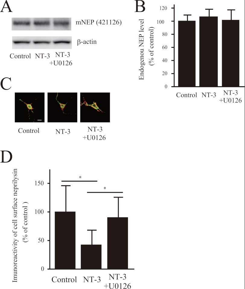
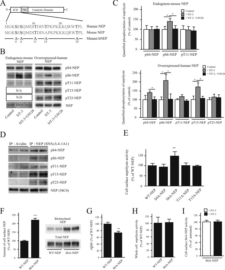
Neprilysin is one of the major amyloid-β peptide (Aβ)-degrading enzymes, the expression of which declines in the brain during aging. The decrease in neprilysin leads to a metabolic Aβ imbalance, which can induce the amyloidosis underlying Alzheimer disease. Pharmacological activation of neprilysin during aging therefore represents a potential strategy to prevent the development of Alzheimer disease. However, the regulatory mechanisms mediating neprilysin activity in the brain remain unclear. To address this issue, we screened for pharmacological regulators of neprilysin activity and found that the neurotrophic factors brain-derived neurotrophic factor, nerve growth factor, and neurotrophins 3 and 4 reduce cell surface neprilysin activity. This decrease was mediated by MEK/ERK signaling, which enhanced phosphorylation at serine 6 in the neprilysin intracellular domain (S6-NEP-ICD). Increased phosphorylation of S6-NEP-ICD in primary neurons reduced the levels of cell surface neprilysin and led to a subsequent increase in extracellular Aβ levels. Furthermore, a specific inhibitor of protein phosphatase-1a, tautomycetin, induced extensive phosphorylation of the S6-NEP-ICD, resulting in reduced cell surface neprilysin activity. In contrast, activation of protein phosphatase-1a increased cell surface neprilysin activity and lowered Aβ levels. Taken together, these results indicate that the phosphorylation status of S6-NEP-ICD influences the localization of neprilysin and affects extracellular Aβ levels. Therefore, maintaining S6-NEP-ICD in a dephosphorylated state, either by inhibition of protein kinases involved in its phosphorylation or by activation of phosphatases catalyzing its dephosphorylation, may represent a new approach to prevent reduction of cell surface neprilysin activity during aging and to maintain physiological levels of Aβ in the brain.
脑啡肽酶是主要的淀粉样β肽(Aβ)降解酶之一,其在大脑中的表达随着年龄的增长而下降。脑啡肽酶的减少导致 Aβ代谢失衡,从而引发阿尔茨海默病的淀粉样变性。因此,在衰老过程中激活脑啡肽酶代表了预防阿尔茨海默病发展的潜在策略。然而,介导大脑中脑啡肽酶活性的调节机制仍不清楚。为了解决这个问题,我们筛选了脑啡肽酶活性的药理学调节剂,发现神经营养因子脑源性神经营养因子、神经生长因子和神经营养因子 3 和 4 降低了细胞表面脑啡肽酶的活性。这种减少是由 MEK/ERK 信号介导的,该信号增强了脑啡肽酶细胞内结构域(S6-NEP-ICD)中丝氨酸 6 的磷酸化。S6-NEP-ICD 的磷酸化增加导致细胞表面脑啡肽酶水平降低,并导致细胞外 Aβ水平随后增加。此外,蛋白磷酸酶-1a 的特异性抑制剂 tautomycetin 诱导 S6-NEP-ICD 的广泛磷酸化,导致细胞表面脑啡肽酶活性降低。相比之下,蛋白磷酸酶-1a 的激活增加了细胞表面脑啡肽酶的活性并降低了 Aβ 水平。总之,这些结果表明 S6-NEP-ICD 的磷酸化状态影响脑啡肽酶的定位,并影响细胞外 Aβ 水平。因此,通过抑制参与其磷酸化的蛋白激酶或通过激活催化其去磷酸化的磷酸酶来维持 S6-NEP-ICD 的去磷酸化状态,可能代表一种新的方法来预防衰老过程中细胞表面脑啡肽酶活性的降低,并维持大脑中 Aβ 的生理水平。