Department of Bio-Nano Science and Engineering, Key Laboratory for Thin Film and Microfabrication of Ministry of Education, Research Institute of Micro/Nano Science and Technology, Shanghai Jiao Tong University, Shanghai200240, P. R. China.
Theranostics. 2012;2(6):618-28. doi: 10.7150/thno.4561. Epub 2012 Jun 15.
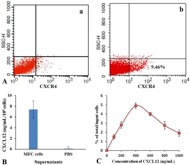
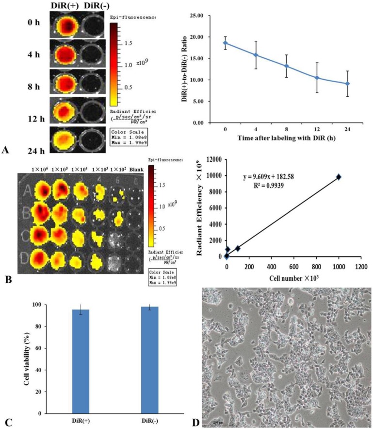
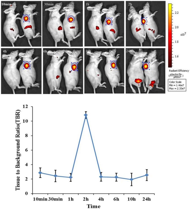
Embryonic stem (ES) cells have great potential in applications such as disease modeling, pharmacological screening and stem cell therapies. Up to date, there is no related report on the use of ES cells as tracking and contrast reagents of cancer cells in vivo. Herein we report that DiR-labeled murine ES cells can recognize and target gastric cancer cells in vivo. DiR-labeled murine ES (mES) cells (5×10(6)) were intravenously injected into gastric tumor-bearing mice. The biodistribution of DiR-labeled mES cells was monitored by IVIS imaging within 24 h. Major organs were harvested and analyzed by immunofluorescence staining and Western blotting. Chemotaxis assay was employed to investigate the chemotaxis of ES cells tracking cancer cells. Fluorescent imaging results showed that DiR-labeled mES cells targeted gastric cancer tissue in vivo as early as 10 min post-injection, reaching a peak at 2h post-injection. Immunofluorescence staining and Western blotting results showed gastric cancer tissues specifically expressed SSEA-1. In vitro migration tests confirmed that mES cells actively moved to test sites with different concentration of CXCL12 in a dose-dependent manner. In conclusion, DiR-labeled mES cells may be used for gastric cancer targeted imaging in vivo, and have great potential in applications such as identifying and imaging of early gastric cancer in near future.
胚胎干细胞(ES 细胞)在疾病建模、药物筛选和干细胞治疗等方面具有巨大的应用潜力。迄今为止,尚无将 ES 细胞用作体内癌细胞跟踪和对比试剂的相关报道。本文报道了 DiR 标记的鼠 ES 细胞可在体内识别和靶向胃癌细胞。将 DiR 标记的鼠 ES(mES)细胞(5×10(6))静脉注射到荷胃癌小鼠体内。在 24 小时内通过 IVIS 成像监测 DiR 标记的 mES 细胞的生物分布。通过免疫荧光染色和 Western blot 分析收集和分析主要器官。采用趋化性测定法研究 ES 细胞追踪癌细胞的趋化性。荧光成像结果表明,DiR 标记的 mES 细胞在注射后 10 分钟即可靶向体内胃癌组织,在注射后 2 小时达到峰值。免疫荧光染色和 Western blot 结果表明胃癌组织特异性表达 SSEA-1。体外迁移试验证实 mES 细胞可主动向不同浓度 CXCL12 的测试部位迁移,呈浓度依赖性。总之,DiR 标记的 mES 细胞可用于体内胃癌的靶向成像,在识别和成像早期胃癌等方面具有广阔的应用前景。