Ruan Jing, Ji Jiajia, Song Hua, Qian Qirong, Wang Kan, Wang Can, Cui Daxiang
Department of Bio-Nano Science and Engineering, Key Laboratory for Thin Film and Microfabrication of Ministry of Education, Institute of Micro-Nano Science and Technology, Shanghai Jiao Tong University, 800 Dongchuan Road, Shanghai, 200240, People's Republic of China.
Nanoscale Res Lett. 2012 Jun 18;7(1):309. doi: 10.1186/1556-276X-7-309.
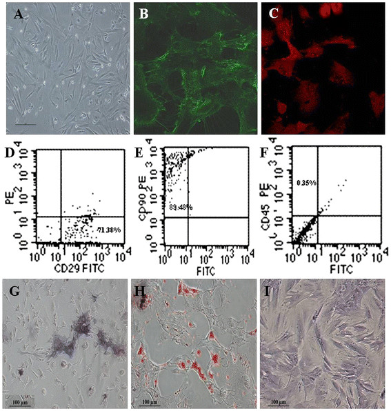
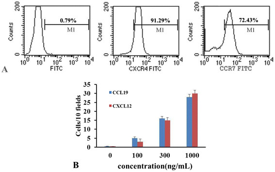
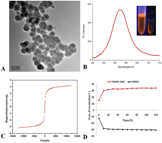
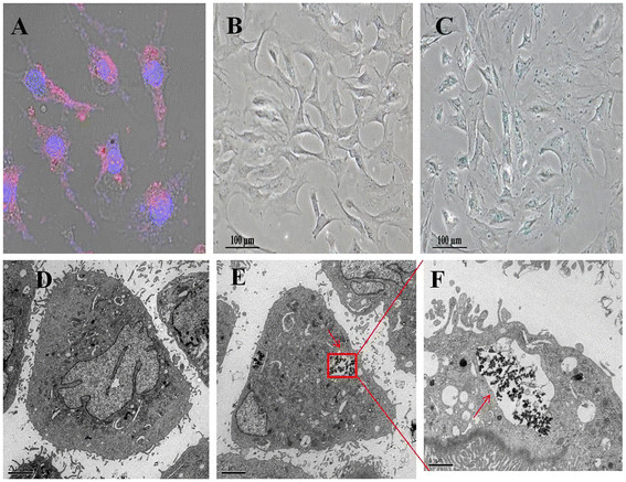
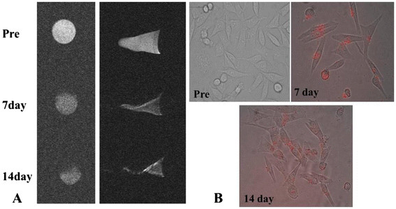
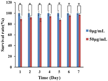
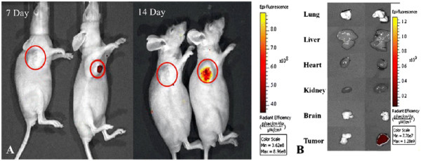
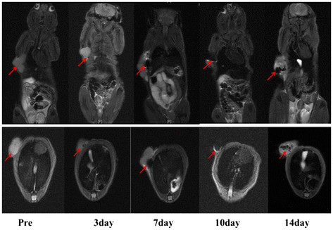
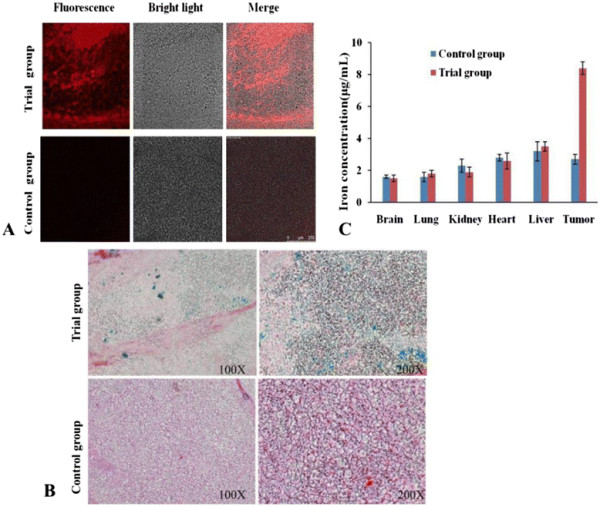
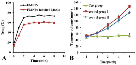
How to find early gastric cancer cells in vivo is a great challenge for the diagnosis and therapy of gastric cancer. This study is aimed at investigating the feasibility of using fluorescent magnetic nanoparticle (FMNP)-labeled mesenchymal stem cells (MSCs) to realize targeted imaging and hyperthermia therapy of in vivo gastric cancer. The primary cultured mouse marrow MSCs were labeled with amino-modified FMNPs then intravenously injected into mouse model with subcutaneous gastric tumor, and then, the in vivo distribution of FMNP-labeled MSCs was observed by using fluorescence imaging system and magnetic resonance imaging system. After FMNP-labeled MSCs arrived in local tumor tissues, subcutaneous tumor tissues in nude mice were treated under external alternating magnetic field. The possible mechanism of MSCs targeting gastric cancer was investigated by using a micro-multiwell chemotaxis chamber assay. Results show that MSCs were labeled with FMNPs efficiently and kept stable fluorescent signal and magnetic properties within 14 days, FMNP-labeled MSCs could target and image in vivo gastric cancer cells after being intravenously injected for 14 days, FMNP-labeled MSCs could significantly inhibit the growth of in vivo gastric cancer because of hyperthermia effects, and CCL19/CCR7 and CXCL12/CXCR4 axis loops may play key roles in the targeting of MSCs to in vivo gastric cancer. In conclusion, FMNP-labeled MSCs could target in vivo gastric cancer cells and have great potential in applications such as imaging, diagnosis, and hyperthermia therapy of early gastric cancer in the near future.
如何在体内发现早期胃癌细胞是胃癌诊断和治疗面临的巨大挑战。本研究旨在探讨使用荧光磁性纳米颗粒(FMNP)标记的间充质干细胞(MSC)实现体内胃癌靶向成像和热疗的可行性。将原代培养的小鼠骨髓间充质干细胞用氨基修饰的FMNP标记,然后静脉注射到皮下胃癌小鼠模型中,随后,利用荧光成像系统和磁共振成像系统观察FMNP标记的间充质干细胞在体内的分布。在FMNP标记的间充质干细胞到达局部肿瘤组织后,对裸鼠的皮下肿瘤组织进行外部交变磁场处理。利用微孔多室趋化性分析方法研究了间充质干细胞靶向胃癌的可能机制。结果表明,间充质干细胞被FMNP有效标记,在14天内保持稳定的荧光信号和磁性,静脉注射14天后,FMNP标记的间充质干细胞能够在体内靶向并成像胃癌细胞,由于热效应,FMNP标记的间充质干细胞能够显著抑制体内胃癌的生长,CCL19/CCR7和CXCL12/CXCR4轴环可能在间充质干细胞对体内胃癌的靶向中起关键作用。总之,FMNP标记的间充质干细胞能够在体内靶向胃癌细胞,在不久的将来,在早期胃癌的成像、诊断和热疗等应用中具有巨大潜力。