NHMRC National Centre of Research Excellence for Chronic Respiratory Disease, School of Medicine, University of Tasmania, Hobart, Australia.
PLoS One. 2012;7(6):e39736. doi: 10.1371/journal.pone.0039736. Epub 2012 Jun 29.
Transforming growth factor-beta1 (TGF-β1) is a multipotential cytokine with angiogenic activity. There are only limited data about its role in airway remodeling in COPD. We have previously shown that the reticular basement membrane (Rbm) is hypervascular in the airways of current smokers either with or without chronic obstructive pulmonary disease (COPD). This study evaluated TGF-β1 immunostaining in the Rbm and its relationship to vascularity in smokers with or without COPD.
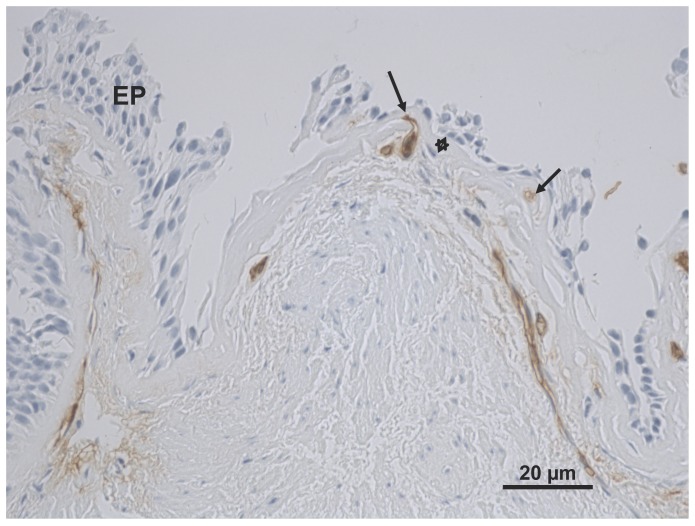
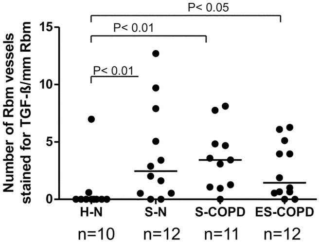
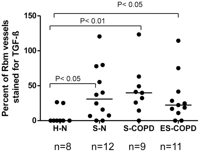
METHODOLOGY/PRINCIPAL FINDINGS: Bronchial biopsies from 15 smokers with normal lung function, 19 current and 14 ex-smokers with COPD were immunostained for TGF-β1 antibody and compared to 17 healthy controls. The percentage area of tissue and also number and area of vessels staining positively for TGF-β1 were measured and compared between groups. Some bronchial biopsies from current smoking COPD subjects were also stained for phosphorylated (active) Smad2/3. Epithelial TGF- β1 staining was not different between COPD current smokers and normal controls. TGF-β1 stained vessels in the Rbm were increased in smokers with normal lung function, current smoking COPD and ex-smokers with COPD compared to controls [median (range) for number of vessels/mm Rbm 2.5 (0.0-12.7), 3.4 (0.0-8.1) and 1.0 (0.0-6.3) vs. 0.0 (0.0-7.0), p<0.05]. Percentage of vessels stained was also increased in these clinical groups. Preliminary data suggest that in current smoking COPD subjects endothelial cells and cells in the Rbm stain positively for phosphorylated Smad2/3 suggesting TGF-β1 is functionally active in this situation.
CONCLUSIONS/SIGNIFICANCE: Vessel-associated TGF-β1 activity is increased in the bronchial Rbm in smokers and especially those with COPD.
转化生长因子-β1(TGF-β1)是一种具有血管生成活性的多功能细胞因子。关于其在 COPD 气道重塑中的作用,仅有有限的数据。我们之前已经表明,在当前吸烟者或患有慢性阻塞性肺疾病(COPD)的吸烟者的气道中,网状基底膜(Rbm)是高血管的。这项研究评估了 TGF-β1 在 Rbm 中的免疫染色及其与吸烟者有无 COPD 之间的血管生成的关系。
方法/主要发现:对 15 名肺功能正常的吸烟者、19 名当前吸烟者和 14 名戒烟者的支气管活检进行 TGF-β1 抗体免疫染色,并与 17 名健康对照组进行比较。测量并比较了组织的阳性染色面积以及阳性染色的血管数量和面积。还对当前吸烟 COPD 患者的一些支气管活检进行了磷酸化(活性)Smad2/3 的染色。COPD 吸烟者和正常对照组之间,上皮细胞 TGF-β1 染色无差异。与对照组相比,肺功能正常的吸烟者、当前吸烟的 COPD 患者和戒烟的 COPD 患者的 Rbm 中 TGF-β1 染色的血管增加[Rbm 中血管数/mm 的中位数(范围)分别为 2.5(0.0-12.7)、3.4(0.0-8.1)和 1.0(0.0-6.3)与 0.0(0.0-7.0)相比,p<0.05]。这些临床组中染色的血管百分比也增加了。初步数据表明,在当前吸烟的 COPD 患者中,内皮细胞和 Rbm 中的细胞对磷酸化 Smad2/3 呈阳性染色,表明 TGF-β1 在这种情况下具有功能活性。
结论/意义:在吸烟者,尤其是 COPD 患者的支气管 Rbm 中,与血管相关的 TGF-β1 活性增加。