Department of Hematology, Singapore General Hospital, Singapore.
Cytotherapy. 2012 Oct;14(9):1064-79. doi: 10.3109/14653249.2012.697146. Epub 2012 Jul 10.
Mesenchymal stromal cells (MSC) have been observed to participate in tissue repair and to have growth-promoting effects on ex vivo co-culture with other stem cells.
In order to evaluate the mechanism of MSC support on ex vivo cultures, we performed co-culture of MSC with umbilical cord blood (UCB) mononuclear cells (MNC) (UCB-MNC).
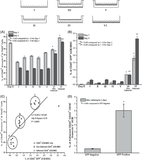
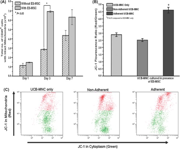
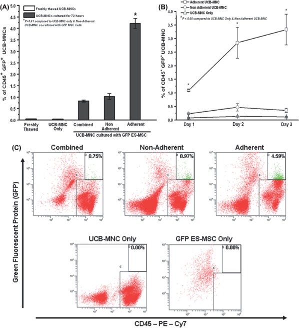
Significant enhancement in cell growth correlating with cell viability was noted with MSC co-culture (defined by double-negative staining for Annexin-V and 7-AAD; P < 0.01). This was associated with significant enhancement of mitochondrial membrane potential (P < 0.01). We postulated that intercellular transfer of cytosolic substances between MSC and UCB-MNC could be one mechanism mediating the support. Using MSC endogenously expressing green fluorescent protein (GFP) or labeled with quantum dots (QD), we performed co-culture of UCB-MNC with these MSC. Transfer of these GFP and QD was observed from MSC to UCB-MNC as early as 24 h post co-culture. Transwell experiments revealed that direct contact between MSC and UCB-MNC was necessary for both transfer and viability support. UCB-MNC tightly adherent to the MSC layer exhibited the most optimal transfer and rescue of cell viability. DNA analysis of the viable, GFP transfer-positive UCB-MNC ruled out MSC transdifferentiation or MSC-UCB fusion. In addition, there was statistical correlation between higher levels of cytosolic transfer and enhanced UCB-MNC viability (P < 0.0001).
Collectively, the data suggest that intercellular transfer of cytosolic materials could be one novel mechanism for preventing UCB cell death in MSC co-culture.
间充质基质细胞(MSC)被观察到参与组织修复,并对与其他干细胞的体外共培养具有促进生长的作用。
为了评估 MSC 对体外培养的支持机制,我们进行了 MSC 与脐带血(UCB)单核细胞(MNC)(UCB-MNC)的共培养。
与 MSC 共培养时,观察到细胞生长显著增强,与细胞活力相关(通过双重阴性染色 Annexin-V 和 7-AAD 定义;P < 0.01)。这与线粒体膜电位的显著增强相关(P < 0.01)。我们推测,MSC 和 UCB-MNC 之间细胞质物质的细胞间转移可能是介导这种支持的一种机制。使用内源性表达绿色荧光蛋白(GFP)或标记量子点(QD)的 MSC,我们进行了 UCB-MNC 与这些 MSC 的共培养。早在共培养后 24 小时,就观察到这些 GFP 和 QD 从 MSC 转移到 UCB-MNC。Transwell 实验表明,MSC 和 UCB-MNC 之间的直接接触对于转移和存活支持都是必要的。紧密附着在 MSC 层上的 UCB-MNC 表现出最佳的转移和细胞活力挽救。对存活、GFP 转移阳性的 UCB-MNC 的 DNA 分析排除了 MSC 转分化或 MSC-UCB 融合。此外,细胞质转移水平与 UCB-MNC 活力的增强之间存在统计学相关性(P < 0.0001)。
综上所述,数据表明细胞质物质的细胞间转移可能是 MSC 共培养中防止 UCB 细胞死亡的一种新机制。