Department of Surgery and Hepatitis Research Center, National Taiwan University Hospital, Taipei, Taiwan.
Int J Nanomedicine. 2012;7:2987-96. doi: 10.2147/IJN.S30061. Epub 2012 Jun 22.
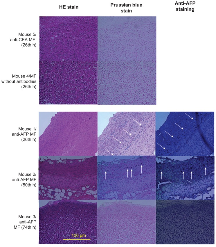
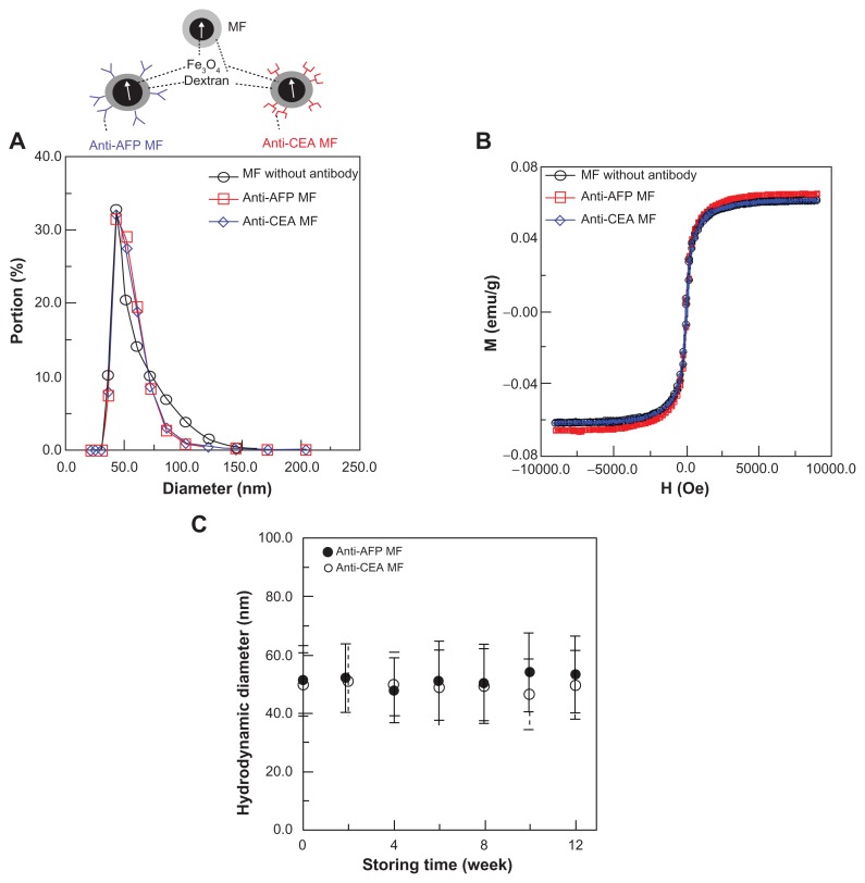
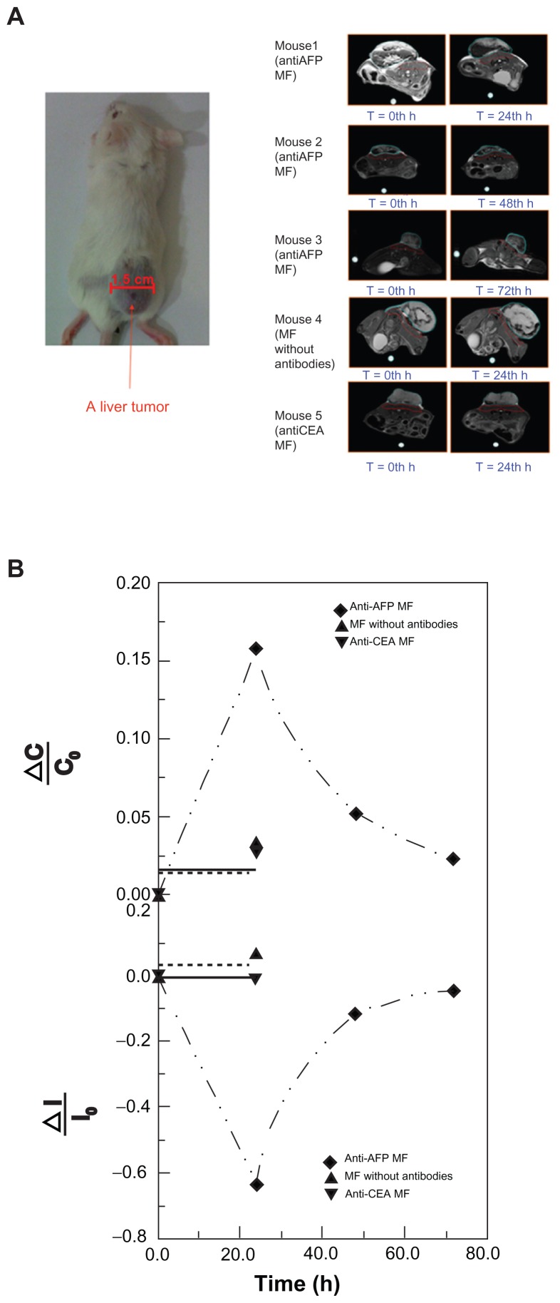
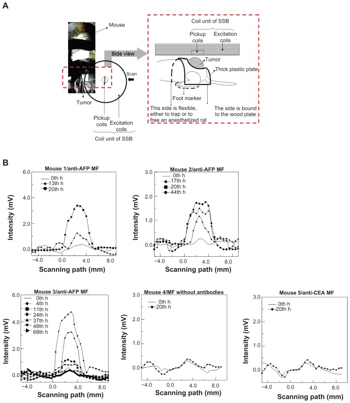
For preoperative and intraoperative detection of tumor distribution, numerous multimodal contrast agents, such as magnetic nanoparticles (MNPs) with several examination indicators, are currently in development. However, complex materials, configuration, and cost are required for multimodal contrast agents, accompanied by a high possibility of toxicity and low popularity in clinics. Nevertheless, the magnetic labeling of MNPs using bioprobes should be feasible not only in preoperative magnetic resonance imaging (MRI), but also in intraoperative examination based on other magnetic properties. In this study, anti-alpha-fetoprotein (AFP)-mediated Fe(3)O(4) MNPs, injected into mice with liver tumors, were used to examine the characteristics of magnetic labeling. Using MRI and scanning superconducting-quantum-interference-device biosusceptometry (SSB), based on alternating current (AC) susceptibility, the magnetic labeling occurred significantly on the first day post-injection of anti-AFP magnetic fluid (MF), and then decreased over time. However, for both MF without antibodies and an anti-carcinoembryonic antigen MF, no magnetic labeling occured on the first day of their respective post-injection. The favorable agreement indicates that magnetic labeling possesses two magnetic characteristics: distortion of the imaging field and AC susceptibility. In addition, the results of the biopsy tests, anti-AFP staining, and Prussian blue staining show the same dynamics as those of magnetic methodologies and prove that bound MNPs on tumor tissue are rotatable by an AC magnetic field to express AC susceptibility. Therefore, with the simple configuration of antibody-mediated MNPs, magnetic labeling is also feasible for intraoperative examinations using SSB with high mobility and sensitivity.
为了在术前和术中检测肿瘤分布,目前正在开发许多多模态造影剂,如具有多种检查指标的磁性纳米颗粒 (MNPs)。然而,多模态造影剂需要复杂的材料、结构和成本,同时伴随着毒性的可能性和在临床上的低普及度。然而,使用生物探针对 MNPs 进行磁性标记不仅在术前磁共振成像 (MRI) 中是可行的,而且在基于其他磁性特性的术中检查中也是可行的。在这项研究中,注射到患有肝肿瘤的小鼠体内的抗甲胎蛋白 (AFP)介导的 Fe(3)O(4) MNPs 被用于检查磁性标记的特征。使用 MRI 和基于交流 (AC) 磁化率的超导量子干涉器件生物磁强计 (SSB),在抗 AFP 磁流体 (MF) 注射后的第一天,磁性标记明显发生,然后随着时间的推移而减少。然而,对于没有抗体的 MF 和抗癌胚抗原 MF,在各自注射后的第一天都没有发生磁性标记。良好的一致性表明,磁性标记具有两种磁性特征:成像场的变形和 AC 磁化率。此外,活检测试、抗 AFP 染色和普鲁士蓝染色的结果与磁方法的结果相同,证明结合在肿瘤组织上的 MNPs 可以通过交流磁场旋转来表达 AC 磁化率。因此,通过抗体介导的 MNPs 的简单配置,磁性标记也可以使用具有高机动性和灵敏度的 SSB 进行术中检查。