• 文献检索
  • 文档翻译
  • 深度研究
  • 学术资讯
  • Suppr Zotero 插件Zotero 插件
  • 邀请有礼
  • 套餐&价格
  • 历史记录
应用&插件
Suppr Zotero 插件Zotero 插件浏览器插件Mac 客户端Windows 客户端微信小程序
定价
高级版会员购买积分包购买API积分包
服务
文献检索文档翻译深度研究API 文档MCP 服务
关于我们
关于 Suppr公司介绍联系我们用户协议隐私条款
关注我们

Suppr 超能文献

核心技术专利:CN118964589B侵权必究
粤ICP备2023148730 号-1Suppr @ 2026

文献检索

告别复杂PubMed语法,用中文像聊天一样搜索,搜遍4000万医学文献。AI智能推荐,让科研检索更轻松。

立即免费搜索

文件翻译

保留排版,准确专业,支持PDF/Word/PPT等文件格式,支持 12+语言互译。

免费翻译文档

深度研究

AI帮你快速写综述,25分钟生成高质量综述,智能提取关键信息,辅助科研写作。

立即免费体验

兔肠道相关淋巴组织穹窿和M细胞窝中表型不同的T细胞亚群。

Phenotypically distinct subpopulations of T cells in domes and M-cell pockets of rabbit gut-associated lymphoid tissues.

作者信息

Ermak T H, Steger H J, Pappo J

机构信息

Cell Biology and Aging Section, VA Medical Center, San Francisco, California.

出版信息

Immunology. 1990 Dec;71(4):530-7.

PMID:2279738
原文链接:https://pmc.ncbi.nlm.nih.gov/articles/PMC1384874/
Abstract

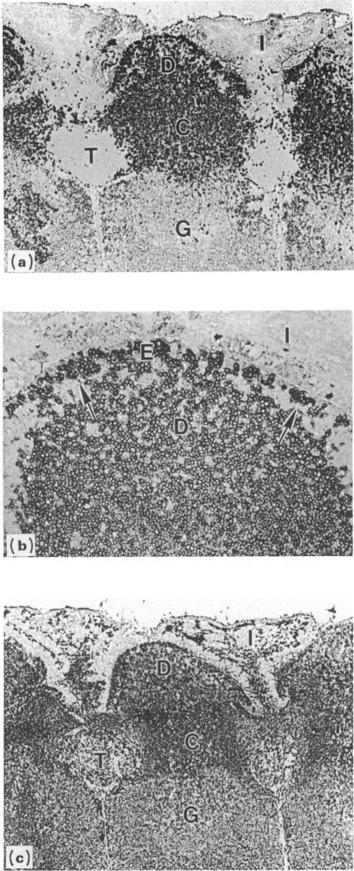
Follicle epithelium and domes of gut-associated lymphoid tissue (GALT) contain populations of lymphocytes which first contact antigen taken up from the intestine. In order to study the association of lymphocytes with M cells in follicle epithelium, monoclonal antibodies (mAb) were generated by immunizing BALB/c mice with lymphocytes populating GALT domes from NZW rabbits, and their specificity was assessed by immunohistochemistry and flow cytometry. mAb 3C10 (IgM) and 3B6 (IgG3) recognized subpopulations of intraepithelial lymphocytes associated with M cells. mAb 3C10 also identified macrophage-lymphocyte clusters in domes and tangible body macrophages in germinal centres of GALT but did not react with cells in T-dependent areas (TDA) or B cells in follicles. mAb 3B6 recognized lymphocytes in domes and B cells in follicles but not T cells in TDA of GALT. The distribution of 3B6+ cells overlapped with, but was more restricted than, that of Ia+ cells. Analysis of lymphocytes in follicle epithelium showed that greater than 95% of lymphocytes associated with M cells were Ia+. T cells represented approximately 95% of intraepithelial lymphocytes in the appendix and approximately 65% in Peyer's patches. A majority of intraepithelial lymphocytes was recognized by mAb 3B6, but mAb 3C10 identified only approximately 30%. Because neither 3C10 nor 3B6 recognized lymphocytes in TDA of GALT, these results indicate that most lymphocytes associated with M cells are a distinct phenotype of Ia+ T cells.

摘要

滤泡上皮和肠道相关淋巴组织(GALT)的圆顶区含有首先接触从肠道摄取的抗原的淋巴细胞群体。为了研究滤泡上皮中淋巴细胞与M细胞的关联,通过用来自新西兰白兔的GALT圆顶区淋巴细胞免疫BALB/c小鼠产生单克隆抗体(mAb),并通过免疫组织化学和流式细胞术评估其特异性。单克隆抗体3C10(IgM)和3B6(IgG3)识别与M细胞相关的上皮内淋巴细胞亚群。单克隆抗体3C10还鉴定了圆顶区的巨噬细胞 - 淋巴细胞簇和GALT生发中心的可触知体巨噬细胞,但不与T细胞依赖区(TDA)中的细胞或滤泡中的B细胞发生反应。单克隆抗体3B6识别圆顶区的淋巴细胞和滤泡中的B细胞,但不识别GALT的TDA中的T细胞。3B6 +细胞的分布与Ia +细胞的分布重叠,但更局限。对滤泡上皮中淋巴细胞的分析表明,与M细胞相关的淋巴细胞中超过95%是Ia +。T细胞约占阑尾上皮内淋巴细胞的95%,在派尔集合淋巴结中约占65%。大多数上皮内淋巴细胞被单克隆抗体3B6识别,但单克隆抗体3C10仅识别约30%。因为3C10和3B6都不识别GALT的TDA中的淋巴细胞,这些结果表明与M细胞相关的大多数淋巴细胞是Ia + T细胞的独特表型。

https://cdn.ncbi.nlm.nih.gov/pmc/blobs/299a/1384874/8b762af4a7ee/immunology00127-0079-a.jpg
https://cdn.ncbi.nlm.nih.gov/pmc/blobs/299a/1384874/4adc99faafd1/immunology00127-0076-a.jpg
https://cdn.ncbi.nlm.nih.gov/pmc/blobs/299a/1384874/5b08a60c7192/immunology00127-0077-a.jpg
https://cdn.ncbi.nlm.nih.gov/pmc/blobs/299a/1384874/6f879d1784b5/immunology00127-0077-b.jpg
https://cdn.ncbi.nlm.nih.gov/pmc/blobs/299a/1384874/d84d002a121a/immunology00127-0078-a.jpg
https://cdn.ncbi.nlm.nih.gov/pmc/blobs/299a/1384874/8b762af4a7ee/immunology00127-0079-a.jpg

相似文献

1
Phenotypically distinct subpopulations of T cells in domes and M-cell pockets of rabbit gut-associated lymphoid tissues.兔肠道相关淋巴组织穹窿和M细胞窝中表型不同的T细胞亚群。
Immunology. 1990 Dec;71(4):530-7.
2
Generation and characterization of monoclonal antibodies recognizing follicle epithelial M cells in rabbit gut-associated lymphoid tissues.识别兔肠道相关淋巴组织中滤泡上皮M细胞的单克隆抗体的产生与特性分析
Cell Immunol. 1989 Apr 15;120(1):31-41. doi: 10.1016/0008-8749(89)90172-x.
3
Mucin-related epitopes distinguish M cells and enterocytes in rabbit appendix and Peyer's patches.黏蛋白相关表位可区分兔阑尾和派尔集合淋巴结中的M细胞和肠上皮细胞。
Infect Immun. 1999 Jan;67(1):357-67. doi: 10.1128/IAI.67.1.357-367.1999.
4
Lymphocyte compartments in antigen-sampling regions of rabbit mucosal lymphoid organs.兔黏膜淋巴器官抗原采样区域的淋巴细胞区室
Am J Trop Med Hyg. 1994;50(5 Suppl):14-28.
5
De novo expression of MECA-79 glycoprotein-determinant on developing B lymphocytes in gut-associated lymphoid tissues.肠道相关淋巴组织中发育中的B淋巴细胞上MECA - 79糖蛋白决定簇的从头表达。
Immunology. 2006 Dec;119(4):461-9. doi: 10.1111/j.1365-2567.2006.02457.x.
6
Heterogeneity of M-cell-associated B and T cells in human Peyer's patches.人派尔集合淋巴结中与微皱褶细胞相关的B细胞和T细胞的异质性。
Immunology. 1994 Nov;83(3):457-64.
7
Experimental mucosal disease in cattle: changes of lymphocyte subpopulations in Peyer's patches and in lymphoid nodules of large intestine.牛的实验性黏膜疾病:派尔集合淋巴结和大肠淋巴小结中淋巴细胞亚群的变化
Vet Immunol Immunopathol. 1995 Oct;48(3-4):233-48. doi: 10.1016/0165-2427(95)05440-h.
8
Large mononuclear Ia-positive veiled cells in Peyer's patches. II. Localization in rat Peyer's patches.派尔集合淋巴结中的大单核Ia阳性被膜细胞。II. 在大鼠派尔集合淋巴结中的定位
Immunology. 1983 Mar;48(3):461-7.
9
Immune cells associated with M cells in the follicle-associated epithelium of Peyer's patches in the rat. An electron- and immuno-electron-microscopic study.大鼠派尔集合淋巴结滤泡相关上皮中与微皱褶细胞相关的免疫细胞。一项电子显微镜和免疫电子显微镜研究。
Cell Tissue Res. 1989 Feb;255(2):293-8. doi: 10.1007/BF00224111.
10
M cells and granular mononuclear cells in Peyer's patch domes of mice depleted of their lymphocytes by total lymphoid irradiation.经全身淋巴照射使淋巴细胞耗竭的小鼠派尔集合淋巴结圆顶区中的M细胞和颗粒单核细胞。
Am J Pathol. 1989 Mar;134(3):529-37.

引用本文的文献

1
The Appendix in Parkinson's Disease: From Vestigial Remnant to Vital Organ?帕金森病中的阑尾:从退化残余到重要器官?
J Parkinsons Dis. 2019;9(s2):S345-S358. doi: 10.3233/JPD-191703.
2
Phenotypic Analysis of a Population of IgA+ Cells in the Follicle-Associated Epithelium of Mouse Peyer's Patches.小鼠派伊尔结滤泡相关上皮中IgA+细胞群体的表型分析
PLoS One. 2015 Apr 20;10(4):e0124111. doi: 10.1371/journal.pone.0124111. eCollection 2015.
3
Unsolved mysteries of intestinal M cells.肠道M细胞的未解之谜。

本文引用的文献

1
Heterogeneity, position, and functional capability of the macrophages in Peyer's patches.派尔集合淋巴结中巨噬细胞的异质性、位置及功能能力。
Cell Tissue Res. 1981;218(3):557-66. doi: 10.1007/BF00210115.
2
Cell renewal and migration in lymphoid follicles of Peyer's patches and cecum--an autoradiographic study in mice.派尔集合淋巴结和盲肠淋巴滤泡中的细胞更新与迁移——小鼠的放射自显影研究
Gastroenterology. 1982 Feb;82(2):232-42.
3
Surface phenotype of Peyer's patch germinal center cells: implications for the role of germinal centers in B cell differentiation.
Gut. 2000 Nov;47(5):735-9. doi: 10.1136/gut.47.5.735.
4
Follicle epithelial M cells are a source of interleukin-1 in Peyer's patches.滤泡上皮M细胞是派尔集合淋巴结中白细胞介素-1的来源。
Immunology. 1993 Mar;78(3):505-7.
5
Uptake and transport of copolymer biodegradable microspheres by rabbit Peyer's patch M cells.兔派伊尔结M细胞对共聚物可生物降解微球的摄取与转运
Cell Tissue Res. 1995 Feb;279(2):433-6. doi: 10.1007/BF00318501.
6
Heterogeneity of M-cell-associated B and T cells in human Peyer's patches.人派尔集合淋巴结中与微皱褶细胞相关的B细胞和T细胞的异质性。
Immunology. 1994 Nov;83(3):457-64.
7
Monoclonal antibody-directed targeting of fluorescent polystyrene microspheres to Peyer's patch M cells.单克隆抗体介导的荧光聚苯乙烯微球靶向派尔集合淋巴结M细胞。
Immunology. 1991 Jul;73(3):277-80.
派尔集合淋巴结生发中心细胞的表面表型:对生发中心在B细胞分化中作用的启示
J Immunol. 1982 Dec;129(6):2698-707.
4
Tissue localization of T and B lymphocytes in lagomorphs: anatomical evidence for a major role of the gastrointestinal associated lymphoid tissue in generation of lymphocytes in the adult.
Dev Comp Immunol. 1980 Spring;4(2):355-66. doi: 10.1016/s0145-305x(80)80038-3.
5
Structure, distribution, and origin of M cells in Peyer's patches of mouse ileum.小鼠回肠派尔集合淋巴结中微皱褶细胞的结构、分布及起源
Gastroenterology. 1984 May;86(5 Pt 1):789-801.
6
Migration of B and T lymphocytes to M cells in Peyer's patch follicle epithelium: an autoradiographic and immunocytochemical study in mice.B淋巴细胞和T淋巴细胞向派尔集合淋巴结滤泡上皮中的M细胞迁移:小鼠的放射自显影和免疫细胞化学研究
Cell Immunol. 1983 Oct 1;81(1):105-17. doi: 10.1016/0008-8749(83)90216-2.
7
Ontogenetic development of T- and B-lymphocytes and non-lymphoid cells in the white pulp of the rat spleen.大鼠脾脏白髓中T淋巴细胞、B淋巴细胞及非淋巴细胞的个体发生发育
Cell Tissue Res. 1983;229(2):351-63. doi: 10.1007/BF00214978.
8
Differentiation antigens identify subpopulations of rabbit T and B lymphocytes. Definition by flow cytometry.分化抗原可识别兔T淋巴细胞和B淋巴细胞的亚群。通过流式细胞术进行定义。
J Exp Med. 1983 Jan 1;157(1):34-46. doi: 10.1084/jem.157.1.34.
9
The anatomy of peripheral lymphoid organs with emphasis on accessory cells: light-microscopic immunocytochemical studies of mouse spleen, lymph node, and Peyer's patch.外周淋巴器官的解剖学,重点是辅助细胞:小鼠脾脏、淋巴结和派尔集合淋巴结的光镜免疫细胞化学研究
Am J Anat. 1984 Jul;170(3):465-81. doi: 10.1002/aja.1001700318.
10
The distribution, ontogeny and origin in the rat of Ia-positive cells with dendritic morphology and of Ia antigen in epithelia, with special reference to the intestine.大鼠中具有树突形态的Ia阳性细胞以及上皮中Ia抗原的分布、个体发生和起源,特别涉及肠道。
Eur J Immunol. 1983 Feb;13(2):112-22. doi: 10.1002/eji.1830130206.