• 文献检索
  • 文档翻译
  • 深度研究
  • 学术资讯
  • Suppr Zotero 插件Zotero 插件
  • 邀请有礼
  • 套餐&价格
  • 历史记录
应用&插件
Suppr Zotero 插件Zotero 插件浏览器插件Mac 客户端Windows 客户端微信小程序
定价
高级版会员购买积分包购买API积分包
服务
文献检索文档翻译深度研究API 文档MCP 服务
关于我们
关于 Suppr公司介绍联系我们用户协议隐私条款
关注我们

Suppr 超能文献

核心技术专利:CN118964589B侵权必究
粤ICP备2023148730 号-1Suppr @ 2026

文献检索

告别复杂PubMed语法,用中文像聊天一样搜索,搜遍4000万医学文献。AI智能推荐,让科研检索更轻松。

立即免费搜索

文件翻译

保留排版,准确专业,支持PDF/Word/PPT等文件格式,支持 12+语言互译。

免费翻译文档

深度研究

AI帮你快速写综述,25分钟生成高质量综述,智能提取关键信息,辅助科研写作。

立即免费体验

经全身淋巴照射使淋巴细胞耗竭的小鼠派尔集合淋巴结圆顶区中的M细胞和颗粒单核细胞。

M cells and granular mononuclear cells in Peyer's patch domes of mice depleted of their lymphocytes by total lymphoid irradiation.

作者信息

Ermak T H, Steger H J, Strober S, Owen R L

机构信息

Department of Medicine, University of California, San Francisco.

出版信息

Am J Pathol. 1989 Mar;134(3):529-37.

PMID:2923183
原文链接:https://pmc.ncbi.nlm.nih.gov/articles/PMC1879537/
Abstract

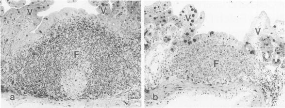
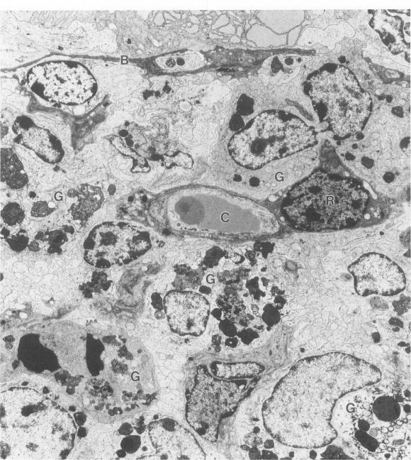
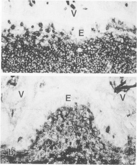
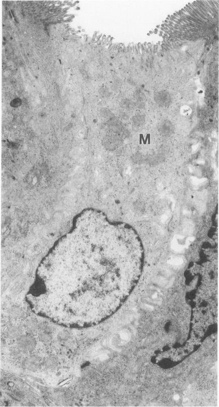
The cytoarchitecture of Peyer's patches that were depleted of their lymphocytes by total lymphoid irradiation (TLI) was examined with particular attention to the effects on M cells in the follicle epithelium and on mononuclear cells in follicle domes underlying the epithelium. Five-month-old, specific pathogen-free Balb/c mice were irradiated with 200-250 rad/day, five times a week to a total dose of 3400-4250, and their Peyer's patches were either fixed for electron microscopy or frozen for immunohistochemistry 1-4 days after completion of irradiation. Control mice were examined at the same time intervals. Follicle domes of TLI mice had approximately one fourth the epithelial surface area of domes of control mice. Within the epithelium, lymphoid cells were virtually depleted after TLI, and yet the epithelium contained M cells. In control mice, most M cells were accompanied by lymphoid cells in invaginations of the apical-lateral cell membrane. In TLI mice, most M cells did not have such apical-lateral invaginations and were columnar shaped. Other than lacking lymphocytes, these cells appeared to be mature M cells. Some M cells did have lymphoid cells or granular mononuclear cells below their basal membranes, adjacent to the basal lamina. Below the epithelium, the proportion of granular mononuclear cells was greatly increased following TLI. The retention of M cells and the increase in proportion of granular mononuclear cells in follicle domes are consistent with selective depletion of lymphocytes following TLI. Persistence of M cells without lymphocytic invaginations after TLI suggests that M cells can differentiate in the absence of, or at least in the presence of very few, lymphocytes, and that invagination by lymphocytes is not necessary to maintain mature M cell morphology.

摘要

通过全淋巴照射(TLI)去除淋巴细胞的派尔集合淋巴结的细胞结构被进行了检查,特别关注其对滤泡上皮中的M细胞以及上皮下方滤泡圆顶中的单核细胞的影响。对5月龄的无特定病原体的Balb/c小鼠,以每天200 - 250拉德的剂量、每周5次进行照射,总剂量达3400 - 4250拉德,在照射完成后1 - 4天,将它们的派尔集合淋巴结要么固定用于电子显微镜检查,要么冷冻用于免疫组织化学检查。同时对对照小鼠进行相同时间间隔的检查。TLI小鼠的滤泡圆顶的上皮表面积约为对照小鼠圆顶的四分之一。在上皮内,TLI后淋巴细胞几乎被耗尽,但上皮中仍含有M细胞。在对照小鼠中,大多数M细胞在顶端 - 侧面细胞膜的内陷处伴有淋巴细胞。在TLI小鼠中,大多数M细胞没有这种顶端 - 侧面内陷,呈柱状。除了缺乏淋巴细胞外,这些细胞似乎是成熟的M细胞。一些M细胞在其基底膜下方、紧邻基膜处确实有淋巴细胞或颗粒状单核细胞。在TLI后,上皮下方颗粒状单核细胞的比例大幅增加。滤泡圆顶中M细胞的保留以及颗粒状单核细胞比例的增加与TLI后淋巴细胞的选择性耗竭一致。TLI后没有淋巴细胞内陷的M细胞的持续存在表明,M细胞可以在没有淋巴细胞的情况下分化,或者至少在淋巴细胞极少的情况下分化,并且淋巴细胞的内陷对于维持成熟M细胞的形态不是必需的。

https://cdn.ncbi.nlm.nih.gov/pmc/blobs/2e57/1879537/64e897869824/amjpathol00123-0053-a.jpg
https://cdn.ncbi.nlm.nih.gov/pmc/blobs/2e57/1879537/f9c9c9dc0e74/amjpathol00123-0048-a.jpg
https://cdn.ncbi.nlm.nih.gov/pmc/blobs/2e57/1879537/5424ac314e43/amjpathol00123-0049-a.jpg
https://cdn.ncbi.nlm.nih.gov/pmc/blobs/2e57/1879537/1f1eb431311c/amjpathol00123-0049-b.jpg
https://cdn.ncbi.nlm.nih.gov/pmc/blobs/2e57/1879537/d70842b50fe5/amjpathol00123-0050-a.jpg
https://cdn.ncbi.nlm.nih.gov/pmc/blobs/2e57/1879537/6ec485e883ea/amjpathol00123-0050-b.jpg
https://cdn.ncbi.nlm.nih.gov/pmc/blobs/2e57/1879537/ddee92c87a1b/amjpathol00123-0051-a.jpg
https://cdn.ncbi.nlm.nih.gov/pmc/blobs/2e57/1879537/77b458382788/amjpathol00123-0052-a.jpg
https://cdn.ncbi.nlm.nih.gov/pmc/blobs/2e57/1879537/64e897869824/amjpathol00123-0053-a.jpg

相似文献

1
M cells and granular mononuclear cells in Peyer's patch domes of mice depleted of their lymphocytes by total lymphoid irradiation.经全身淋巴照射使淋巴细胞耗竭的小鼠派尔集合淋巴结圆顶区中的M细胞和颗粒单核细胞。
Am J Pathol. 1989 Mar;134(3):529-37.
2
Depletion and repopulation of lymphocytes in Peyer's patches of mice after total lymphoid irradiation.全淋巴照射后小鼠派尔集合淋巴结中淋巴细胞的耗竭与再填充。
Lab Invest. 1988 Nov;59(5):591-7.
3
The development of M cells in Peyer's patches is restricted to specialized dome-associated crypts.派尔集合淋巴结中M细胞的发育局限于特化的圆顶相关隐窝。
Am J Pathol. 1999 May;154(5):1573-82. doi: 10.1016/S0002-9440(10)65410-7.
4
Sequential uptake of horseradish peroxidase by lymphoid follicle epithelium of Peyer's patches in the normal unobstructed mouse intestine: an ultrastructural study.正常无梗阻小鼠肠道派尔集合淋巴结淋巴滤泡上皮对辣根过氧化物酶的顺序摄取:一项超微结构研究
Gastroenterology. 1977 Mar;72(3):440-51.
5
Bombesin prevents the atrophy of Peyer's patches and the dysfunction of M cells in rabbits receiving long-term parenteral nutrition.蛙皮素可预防长期接受肠外营养的家兔派尔集合淋巴结萎缩及M细胞功能障碍。
JPEN J Parenter Enteral Nutr. 2007 Mar-Apr;31(2):75-85. doi: 10.1177/014860710703100275.
6
Structure, distribution, and origin of M cells in Peyer's patches of mouse ileum.小鼠回肠派尔集合淋巴结中微皱褶细胞的结构、分布及起源
Gastroenterology. 1984 May;86(5 Pt 1):789-801.
7
Postnatal development of lymphoid follicles in rat Peyer's patches, with special reference to increased follicle number.大鼠派尔集合淋巴结中淋巴滤泡的产后发育,特别提及滤泡数量的增加
Arch Histol Cytol. 1995 Aug;58(3):335-43. doi: 10.1679/aohc.58.335.
8
Salmonella typhimurium initiates murine infection by penetrating and destroying the specialized epithelial M cells of the Peyer's patches.鼠伤寒沙门氏菌通过穿透并破坏派尔集合淋巴结的特化上皮M细胞引发小鼠感染。
J Exp Med. 1994 Jul 1;180(1):15-23. doi: 10.1084/jem.180.1.15.
9
Follicle associated epithelium of the gut associated lymphoid tissue of cattle.牛肠道相关淋巴组织的滤泡相关上皮
Vet Pathol. 1991 Jan;28(1):22-9. doi: 10.1177/030098589102800104.
10
Immunohistochemical localisation of intercellular adhesion molecule-1 in follicle associated epithelium of Peyer's patches.派尔集合淋巴结滤泡相关上皮中细胞间黏附分子-1的免疫组织化学定位
Gut. 1994 Jan;35(1):46-50. doi: 10.1136/gut.35.1.46.

引用本文的文献

1
Unsolved mysteries of intestinal M cells.肠道M细胞的未解之谜。
Gut. 2000 Nov;47(5):735-9. doi: 10.1136/gut.47.5.735.
2
The immunopathology of M cells.M细胞的免疫病理学
Springer Semin Immunopathol. 1997;18(4):421-48. doi: 10.1007/BF00824051.
3
Chloride conductance and intracellular chloride accumulation in mouse Peyer's patch enterocytes.小鼠派尔集合淋巴结肠上皮细胞中的氯电导和细胞内氯蓄积

本文引用的文献

1
"M" cell distribution in follicle-associated epithelium of mouse Peyer's patch.小鼠派尔集合淋巴结滤泡相关上皮中M细胞的分布
Am J Anat. 1980 Oct;159(2):167-75. doi: 10.1002/aja.1001590205.
2
Cell proliferation in follicle-associated epithelium of mouse Peyer's patch.小鼠派尔集合淋巴结滤泡相关上皮中的细胞增殖
Am J Anat. 1980 Oct;159(2):157-66. doi: 10.1002/aja.1001590204.
3
Phagocytosis of Giardia muris by macrophages in Peyer's patch epithelium in mice.小鼠派尔集合淋巴结上皮中巨噬细胞对鼠贾第虫的吞噬作用。
J Physiol. 1990 Aug;427:71-80. doi: 10.1113/jphysiol.1990.sp018161.
4
Structural specializations for antigen uptake and processing in the digestive tract.消化道中抗原摄取和处理的结构特化。
Springer Semin Immunopathol. 1990;12(2-3):139-52. doi: 10.1007/BF00197502.
Infect Immun. 1981 Aug;33(2):591-601. doi: 10.1128/iai.33.2.591-601.1981.
4
Heterogeneity, position, and functional capability of the macrophages in Peyer's patches.派尔集合淋巴结中巨噬细胞的异质性、位置及功能能力。
Cell Tissue Res. 1981;218(3):557-66. doi: 10.1007/BF00210115.
5
Cell renewal and migration in lymphoid follicles of Peyer's patches and cecum--an autoradiographic study in mice.派尔集合淋巴结和盲肠淋巴滤泡中的细胞更新与迁移——小鼠的放射自显影研究
Gastroenterology. 1982 Feb;82(2):232-42.
6
Microcirculation of intestinal lymphoid follicles in rat Peyer's patches.大鼠派尔集合淋巴结中肠淋巴滤泡的微循环
Gastroenterology. 1981 Sep;81(3):481-91.
7
'Managing' the immune system with total lymphoid irradiation.
Hosp Pract (Off Ed). 1981 Jun;16(6):77-89. doi: 10.1080/21548331.1981.11946785.
8
Structure, distribution, and origin of M cells in Peyer's patches of mouse ileum.小鼠回肠派尔集合淋巴结中微皱褶细胞的结构、分布及起源
Gastroenterology. 1984 May;86(5 Pt 1):789-801.
9
Cytochemical analysis of alkaline phosphatase and esterase activities and of lectin-binding and anionic sites in rat and mouse Peyer's patch M cells.大鼠和小鼠派尔集合淋巴结M细胞中碱性磷酸酶和酯酶活性、凝集素结合及阴离子位点的细胞化学分析
Am J Anat. 1983 Oct;168(2):199-212. doi: 10.1002/aja.1001680207.
10
Migration of B and T lymphocytes to M cells in Peyer's patch follicle epithelium: an autoradiographic and immunocytochemical study in mice.B淋巴细胞和T淋巴细胞向派尔集合淋巴结滤泡上皮中的M细胞迁移:小鼠的放射自显影和免疫细胞化学研究
Cell Immunol. 1983 Oct 1;81(1):105-17. doi: 10.1016/0008-8749(83)90216-2.