• 文献检索
  • 文档翻译
  • 深度研究
  • 学术资讯
  • Suppr Zotero 插件Zotero 插件
  • 邀请有礼
  • 套餐&价格
  • 历史记录
应用&插件
Suppr Zotero 插件Zotero 插件浏览器插件Mac 客户端Windows 客户端微信小程序
定价
高级版会员购买积分包购买API积分包
服务
文献检索文档翻译深度研究API 文档MCP 服务
关于我们
关于 Suppr公司介绍联系我们用户协议隐私条款
关注我们

Suppr 超能文献

核心技术专利:CN118964589B侵权必究
粤ICP备2023148730 号-1Suppr @ 2026

文献检索

告别复杂PubMed语法,用中文像聊天一样搜索,搜遍4000万医学文献。AI智能推荐,让科研检索更轻松。

立即免费搜索

文件翻译

保留排版,准确专业,支持PDF/Word/PPT等文件格式,支持 12+语言互译。

免费翻译文档

深度研究

AI帮你快速写综述,25分钟生成高质量综述,智能提取关键信息,辅助科研写作。

立即免费体验

脑桥梗死后小脑中间脚华勒氏变性中的扩散受限

Restricted diffusion in Wallerian degeneration of the middle cerebellar peduncles following pontine infarction.

作者信息

Musson Rachel, Romanowski Charles

机构信息

Department of Radiology, Royal Hallamshire Hospital, Sheffield, U.K.

出版信息

Pol J Radiol. 2010 Oct;75(4):38-43.

PMID:22802803
原文链接:https://pmc.ncbi.nlm.nih.gov/articles/PMC3389899/
Abstract

BACKGROUND

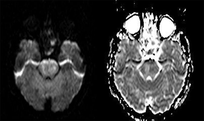
Acute Wallerian degeneration following infarction has been show to result in areas of restricted diffusion within the brain. Very few reports describe this appearance in middle cerebellar peduncles.

CASE REPORT

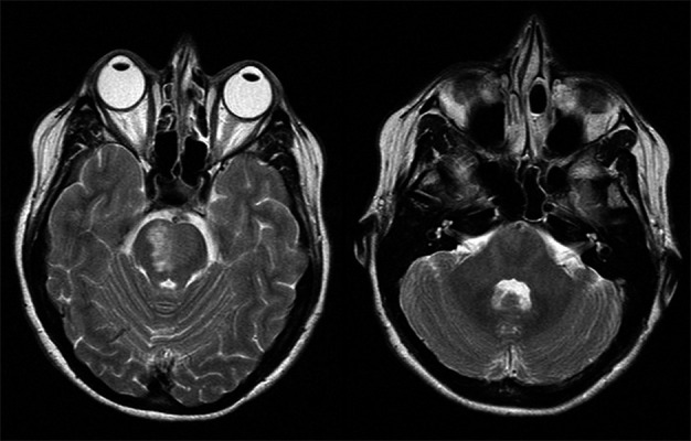
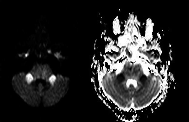
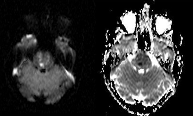
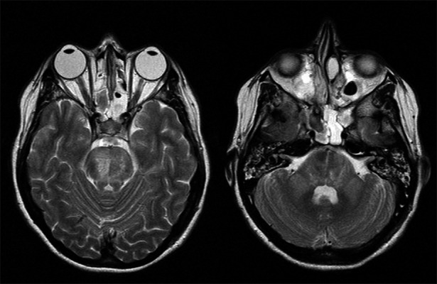
A 37 year old woman was admitted to hospital following sudden collapse and was subsequently found to have a pontine infarct. The complex clinical course resulted in MR imaging including DWI at day 4, 9 and 23 after the initial presentation. The cerebellar peduncles were normal when imaged on day 4 and 9. At day 23, symmetrical high T2 signal was seen in both cerebellar peduncles. The DWI illustrated high signal with corresponding low signal on the ADC map consistent with restricted diffusion. We discuss why this appearance is in keeping with Wallerian degeneration and also describe the fibre pathways involved.

CONCLUSIONS

Symmetrical high signal with restricted diffusion in the middle cerebellar peduncles following a pontine lesion is almost certainly attributable to Wallerian degeneration and should not be mistaken for a new ischaemia.

摘要

背景

梗死继发的急性华勒氏变性已被证实可导致脑内出现扩散受限区域。很少有报道描述小脑中脚出现这种表现。

病例报告

一名37岁女性在突然晕倒后入院,随后被发现患有脑桥梗死。复杂的临床病程导致在首次发病后的第4天、第9天和第23天进行了包括弥散加权成像(DWI)在内的磁共振成像检查。在第4天和第9天成像时,小脑脚正常。在第23天,双侧小脑脚均可见对称的高T2信号。DWI显示高信号,表观扩散系数(ADC)图上相应为低信号,符合扩散受限。我们讨论了为什么这种表现符合华勒氏变性,并描述了相关的纤维束。

结论

脑桥病变后小脑中脚出现对称的高信号且伴有扩散受限,几乎肯定是由华勒氏变性引起的,不应误诊为新的缺血性病变。

https://cdn.ncbi.nlm.nih.gov/pmc/blobs/bd58/3389899/987227d3ad92/poljradiol-75-4-38-g005.jpg
https://cdn.ncbi.nlm.nih.gov/pmc/blobs/bd58/3389899/d491b5d67ece/poljradiol-75-4-38-g001.jpg
https://cdn.ncbi.nlm.nih.gov/pmc/blobs/bd58/3389899/e52c98c103d2/poljradiol-75-4-38-g002.jpg
https://cdn.ncbi.nlm.nih.gov/pmc/blobs/bd58/3389899/e1c8a594a77d/poljradiol-75-4-38-g003.jpg
https://cdn.ncbi.nlm.nih.gov/pmc/blobs/bd58/3389899/93cdd02fd84c/poljradiol-75-4-38-g004.jpg
https://cdn.ncbi.nlm.nih.gov/pmc/blobs/bd58/3389899/987227d3ad92/poljradiol-75-4-38-g005.jpg

相似文献

1
Restricted diffusion in Wallerian degeneration of the middle cerebellar peduncles following pontine infarction.脑桥梗死后小脑中间脚华勒氏变性中的扩散受限
Pol J Radiol. 2010 Oct;75(4):38-43.
2
Neuroimaging and clinical features of bilateral Wallerian degeneration of middle cerebellar peduncles subsequent to pontine infarction.桥脑梗死后继发双侧小脑中脚的沃勒氏变性的神经影像学和临床特征。
CNS Neurosci Ther. 2024 Jul;30(7):e14828. doi: 10.1111/cns.14828.
3
Bilateral wallerian degeneration of the middle cerebellar peduncles secondary to pontine infarction: A case series.桥脑梗死所致的小脑中脚双侧 Wallerian 变性:病例系列研究。
J Neurol Sci. 2018 May 15;388:182-185. doi: 10.1016/j.jns.2018.03.027. Epub 2018 Mar 20.
4
Wallerian degeneration of the middle cerebellar peduncle after pontine infarction: MR imaging.脑桥梗死后小脑中脚的华勒氏变性:磁共振成像
Radiat Med. 2004 Jan-Feb;22(1):37-41.
5
Wallerian degeneration of the ipsilateral middle cerebellar peduncle after lower pontine paramedian infarct diagnosed by magnetic resonance imaging.经磁共振成像诊断为下桥脑旁正中梗塞后同侧小脑中脚的华勒氏变性。
Radiol Case Rep. 2023 Jun 8;18(8):2823-2826. doi: 10.1016/j.radcr.2023.05.033. eCollection 2023 Aug.
6
Clinical and Radiological Features of Wallerian Degeneration of the Middle Cerebellar Peduncles Secondary to Pontine Infarction.桥脑梗死继发的中脑小脑脚瓦勒氏变性的临床和放射学特征。
Chin Med J (Engl). 2018 Mar 20;131(6):665-671. doi: 10.4103/0366-6999.226890.
7
Bilateral Wallerian degeneration of the middle cerebellar peduncles due to paramedian pontine infarction: MRI findings.桥脑旁正中梗死所致双侧小脑中脚华勒氏变性:MRI表现
Neuroradiology. 2004 Nov;46(11):896-9. doi: 10.1007/s00234-004-1287-y. Epub 2004 Oct 29.
8
Bilateral Wallerian Degeneration of the Pontocerebellar Tracts.桥小脑束的双侧华勒氏变性
Case Rep Emerg Med. 2015;2015:970570. doi: 10.1155/2015/970570. Epub 2015 Jun 10.
9
[A case showing Wallerian degeneration of the bilateral middle cerebellar peduncles on MRI followed by the right pontine infarction].[一例MRI显示双侧小脑中脚华勒氏变性,随后发生右侧脑桥梗死的病例]
Rinsho Shinkeigaku. 2004 Feb;44(2):105-7.
10
Bilateral wallerian degeneration of the middle cerebellar peduncles due to unilateral pontine infarction.单侧脑桥梗死导致双侧小脑中脚华勒氏变性。
J Neuroradiol. 2006 Oct;33(4):263-5. doi: 10.1016/s0150-9861(06)77273-x.

引用本文的文献

1
Neuroimaging and clinical features of bilateral Wallerian degeneration of middle cerebellar peduncles subsequent to pontine infarction.桥脑梗死后继发双侧小脑中脚的沃勒氏变性的神经影像学和临床特征。
CNS Neurosci Ther. 2024 Jul;30(7):e14828. doi: 10.1111/cns.14828.
2
Wallerian degeneration of the ipsilateral middle cerebellar peduncle after lower pontine paramedian infarct diagnosed by magnetic resonance imaging.经磁共振成像诊断为下桥脑旁正中梗塞后同侧小脑中脚的华勒氏变性。
Radiol Case Rep. 2023 Jun 8;18(8):2823-2826. doi: 10.1016/j.radcr.2023.05.033. eCollection 2023 Aug.
3
Wallerian degeneration in the brain after organophosphorus poisoning: a case report.

本文引用的文献

1
Progression of pathological changes in the middle cerebellar peduncle by diffusion tensor imaging correlates with lesser motor gains after pontine infarction.通过扩散张量成像观察到的小脑中脚病理变化进展与脑桥梗死后较小的运动功能改善相关。
Neurorehabil Neural Repair. 2009 Sep;23(7):692-8. doi: 10.1177/1545968308331142. Epub 2009 Feb 25.
2
Restricted diffusion in the splenium of the corpus callosum after cardiac arrest.心脏骤停后胼胝体压部的扩散受限
Open Neuroimag J. 2008;2:1-4. doi: 10.2174/1874440000802010001. Epub 2008 Jan 11.
3
[Wallerian degeneration after stroke: a new prognostic factor?].
有机磷中毒后脑内的华勒氏变性:一例报告
Ann Med Surg (Lond). 2023 Mar 27;85(4):926-930. doi: 10.1097/MS9.0000000000000102. eCollection 2023 Apr.
4
Delayed Diffusion Restriction of Wallerian Degeneration.沃勒变性的延迟扩散受限
Neurocrit Care. 2023 Jun;38(3):825-828. doi: 10.1007/s12028-023-01692-7. Epub 2023 Feb 24.
5
Clinical applications of diffusion-weighted sequence in brain imaging: beyond stroke.扩散加权序列在脑成像中的临床应用:超越中风。
Neuroradiology. 2022 Jan;64(1):15-30. doi: 10.1007/s00234-021-02819-3. Epub 2021 Oct 1.
6
Magnetic resonance imaging evaluation of Wallerian degeneration of bilateral middle cerebellar peduncles after pontine infarction.脑桥梗死后双侧小脑中脚华勒氏变性的磁共振成像评估
Saudi Med J. 2019 Dec;40(12):1278-1284. doi: 10.15537/smj.2019.12.24491.
7
Clinical and Radiological Features of Wallerian Degeneration of the Middle Cerebellar Peduncles Secondary to Pontine Infarction.桥脑梗死继发的中脑小脑脚瓦勒氏变性的临床和放射学特征。
Chin Med J (Engl). 2018 Mar 20;131(6):665-671. doi: 10.4103/0366-6999.226890.
8
Extensive Restricted Diffusion in Bilateral Hemispheric White Matter Following Diffuse Hypoxic Injury Due to Hanging.绞缢所致弥漫性缺氧性损伤后双侧半球白质广泛的扩散受限
J Neurosci Rural Pract. 2017 Apr-Jun;8(2):306-307. doi: 10.4103/0976-3147.203841.
9
Middle cerebellar peduncles: Magnetic resonance imaging and pathophysiologic correlate.小脑中脚:磁共振成像与病理生理关联
World J Radiol. 2015 Dec 28;7(12):438-47. doi: 10.4329/wjr.v7.i12.438.
10
Motor pathway degeneration in young ataxia telangiectasia patients: A diffusion tractography study.年轻共济失调毛细血管扩张症患者的运动通路退变:一项扩散张量成像研究。
Neuroimage Clin. 2015 Aug 20;9:206-15. doi: 10.1016/j.nicl.2015.08.007. eCollection 2015.
[中风后沃勒变性:一种新的预后因素?]
Acta Med Port. 2006 Nov-Dec;19(6):499-502. Epub 2007 May 14.
4
Bilateral wallerian degeneration of the middle cerebellar peduncles due to unilateral pontine infarction.单侧脑桥梗死导致双侧小脑中脚华勒氏变性。
J Neuroradiol. 2006 Oct;33(4):263-5. doi: 10.1016/s0150-9861(06)77273-x.
5
Wallerian degeneration of the pontocerebellar fibers.脑桥小脑纤维的华勒氏变性。
AJNR Am J Neuroradiol. 2005 May;26(5):1062-5.
6
Bilateral Wallerian degeneration of the middle cerebellar peduncles due to paramedian pontine infarction: MRI findings.桥脑旁正中梗死所致双侧小脑中脚华勒氏变性:MRI表现
Neuroradiology. 2004 Nov;46(11):896-9. doi: 10.1007/s00234-004-1287-y. Epub 2004 Oct 29.
7
Diffusion tensor imaging detects early Wallerian degeneration of the pyramidal tract after ischemic stroke.弥散张量成像可检测缺血性卒中后锥体束的早期华勒氏变性。
Neuroimage. 2004 Aug;22(4):1767-74. doi: 10.1016/j.neuroimage.2004.03.041.
8
Wallerian degeneration of the middle cerebellar peduncle after pontine infarction: MR imaging.脑桥梗死后小脑中脚的华勒氏变性:磁共振成像
Radiat Med. 2004 Jan-Feb;22(1):37-41.
9
Transient detection of early wallerian degeneration on diffusion-weighted MRI after an acute cerebrovascular accident.急性脑血管意外后弥散加权磁共振成像上早期沃勒变性的短暂性检测
Neuroradiology. 2004 Mar;46(3):183-8. doi: 10.1007/s00234-003-1159-x. Epub 2004 Feb 27.
10
Bilateral Wallerian degeneration of the medial cerebellar peduncles after ponto-mesencephalic infarction.桥中脑梗死继发双侧小脑中脚华勒氏变性
Eur J Radiol. 2004 Mar;49(3):198-203. doi: 10.1016/S0720-048X(03)00132-3.